Webinar
Improve Medical Image Processing — 5 Motives and Methods
About this webinar
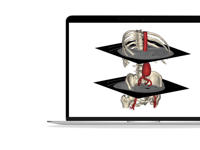
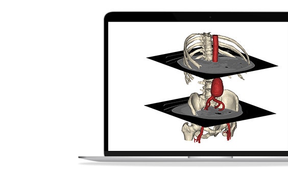
Materialise’s Mimics Innovation Suite can assist users in avoiding setbacks and delays within the design phases. Medical device innovators want to ensure accuracy coming from 3D-CT and MRI when creating surface models. The critical image segmentation techniques outlined in the webinar can be used to create and understand anatomy — all within Materialise’s industry-standard 3D software.
What you will learn
- Ensure accuracy with surface models — discover methods to potentially reduce changes in surgical plans
- Best practices for making R&D deadlines more achievable — bring software solutions in-house to keep your timeline on schedule
- Techniques for cost-efficient anatomical analysis — go beyond basic measurements with center lines, global head maps, and more
- Simplify complex anatomy — surgeons and support staff can use Mimics to create 3D-printed models for better patient communication
- Engage with stable, reliable, and certified tools — Materialise, as a medical device company, understands the unique needs of medtech innovators
Speakers

Maya Dbaibo

Dominic Rinna
Share on:
Materialise medical device software may not be available in all markets because product availability is subject to the regulatory or medical practices in individual markets. In countries where no regulatory registration is obtained of Mimics or 3-matic Medical, a research version is available. Please contact your Materialise representative if you have questions about the availability of Materialise medical device software in your area.
L-103967-01
Scaling Medical Device Design and Commercialization
Leverage Mimics Innovation Suite's tools to improve the quality of your medical device processes. This series of short webinars is designed for medical device companies in every stage of development and is especially relevant for R&D, clinical engineers, product managers, and anyone looking to commercialize patient-specific medical devices using 3D imaging data.