Webinar
3D and Musculoskeletal Trauma
About this webinar
Professor Stefaan Nijs elaborates on how 3D technologies are transforming the field of trauma surgery and improving the quality of life of dozens of patients, many with severe complications that made them untreatable with conventional methods.
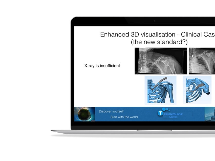
During the webinar, Prof. Nijs demonstrates the capabilities of 3D imaging for accurate diagnosis by presenting a case study of a patient with a calcaneal fracture. 3D visualization is also extremely useful for diagnosis and predicting the best medical procedure. This is exemplified by a case example of a 17-year-old girl who suffered severe injuries after a horseback riding accident. Dr. Nijs concludes with the benefits and reasons why he feels that 3D visualization and guidance make trauma surgery a craft and no longer an art.
What you will learn
- The difference between 2D and 3D images and concrete examples of why 3D images influence the surgical strategy
- How Dr. Nijs uses 3D images and planning pre-, intra- and post-operatively
- What SurgiCase is and how the platform can be used to prepare for surgery
- Case stories and practical examples, illustrated by pictures and videos of how Dr. Nijs uses the 3D technique in his daily practice
- The four benefits of incorporating 3D software technology and 3D printing in the surgical workflow, according to Dr. Nijs
Speaker

Professor Dr. Stefaan Nijs
Share on: