PODCAST
Medical 3D Players: Precision in Practice — The Technology Behind Thoracic Surgery (S2, Ep.03)
Discussing mass personalization in healthcare — because one size fits no one
- Subscribe:
- Apple Podcasts
- |
- YouTube
- |
- Spotify
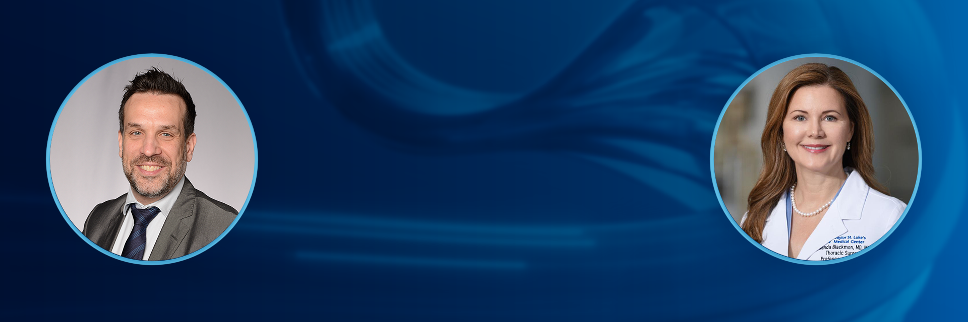
Uncover the latest medical advancements and challenges in 3D technology. Hosted by Pieter Slagmolen and Sebastian De Boodt from Materialise, this podcast examines key developments with experts in the healthcare industry.
In this episode, we sit down with Dr. Shanda H Blackmon, Executive Director of Baylor College of Medicine Lung Institute, and Dr. Igor Saftic, Clinical Director of Thoracic Surgery at University Hospitals Bristol and Weston NHS Foundation Trust, to explore how technology such as 3D planning and robots support thoracic surgery. Don’t miss their insightful call-out on what’s still needed to achieve greater precision and transform patient care in lung surgery.
We'll discuss the investigational use of medical devices. In that context, our guests will provide their own opinions and not medical advice.
- Subscribe:
- Apple Podcasts
- |
- YouTube
- |
- Spotify
Read the full transcript
Pieter Slagmolen 0:06
Welcome to another episode of the 3D Players Podcast, where we explore the intersection of innovation and healthcare. I'm your host, Peter Slagmolen, and I'm joined by my cohost Sebastian De Boodt.
Sebastian De Boodt 0:16
Today, we explore new frontiers of personalization and 3D planning in lung cancer surgery together with two remarkable guests; of course, our first guest is Dr. Igor Saftic, who is clinical director of thoracic surgery at University Hospital Bristol in the UK. Our second guest is Dr. Shanda Blackmon, who is the executive director at Baylor College of Medicine Lung Institute in Houston, Texas. Next to fulfilling leadership functions at their respective institutes, they both are internationally recognized lung surgeons with extensive experience in 3D planning. Welcome, Dr. Saftic and Dr. Blackmon.
Dr. Shanda H Blackmon 0:55
Thank you. It's an honor to be with you.
Pieter Slagmolen 0:57
Dr. Saftic, so let's start with you. Could you share a little bit about your your background and what led you to specialize in lung surgery in specific?
Dr. Igor Saftic 1:10
Okay, hello, and first of all, thank you for having me. So my surgical training, well, I was all over the world, I would say. So most of it was done back in Croatia, where I'm from. I've had training opportunities in Houston, Texas, mainly Texas Heart Methodist and afterward, in Paris and Guys Hospital London, after, which then I've accepted the position of a consultant thoracic surgeon in Bristol, UK, for which I've been a director now for a year. What led me to thoracic surgery? Specifically, I'm afraid I can't really answer this in one sentence. I would say my career started, or my training started in cardiac surgery. And then, I just followed my gut feeling that thoracic surgery would be something that would be quite significantly changing and developing in the future. And yeah, that's that. That's potentially why. So I can satisfy the curiosities I had back then, and I'm still happy I made a choice.
Sebastian De Boodt 2:12
All right, that's good to hear. And Dr. Blackmon, you've also had a fascinating career. Could you tell us what drew you into this field, your journey, and how it has evolved over the years?
Dr. Shanda H Blackmon 2:24
Yes, so Igor and I might have crossed paths at many points and times. I came to Houston, Texas for cardiothoracic surgical training as the primary focus on aortic surgery under Joseph Caselli, and when I went on maternity leave to give birth to my twins, I came back to Houston Methodist Hospital and Texas Heart. Well, Houston Methodist Hospital and Baylor College of Medicine had gone through a bit of a divorce, and I found myself suddenly transplanted over to Texas Heart Hospital and Baylor College of Medicine, what is today, Baylor College of Medicine, St Luke's Episcopal Hospital, now Common Spirit. So when I came over to the new institution, traditionally training in a place that was dominated by Debakey and now a new institution dominated by Denton Cooley, you got to see two giants training one person, and I might have been one of the only people that really got to train under both of those giants for cardiac surgery. And in spite of that, I became also inspired to pursue a career in thoracic surgery. So I did a complex thoracic oncology fellowship at MD Anderson and then and the Founding Chair of the division of thoracic surgery at Houston Methodist Hospital, where I served there for eight years. I was then recruited to go to Mayo Clinic, where I was the medical director for consumer digital platform and the thoracic champion for the 3D printing lab that was initially begun by Jane Matsumoto and then carried on by Jonathan Morris. Finally, I got recruited to come back to Houston, where we have family and old friends and much warmer weather, and I'm practicing thoracic surgical oncology, minimally invasive thoracic surgery, and leading the Lung Institute, and really happy to be back at Baylor College of Medicine, where I did all of my training.
Sebastian De Boodt 4:29
All right, it's good to hear how, by coincidence, you know your paths are now, again crossing on this podcast because you haven't met each other before, right? So it's also, again, you know, a new introduction. Okay, cool,
Pieter Slagmolen 4:44
Maybe as a follow-up question on that, both of you described that you had a history also in cardiac surgery and now evolved into thoracic surgery. If you look at thoracic surgery, what are the types of patients that you're treating there typically? What ailments are you guys trying to address in your day-to-day work?
Dr. Igor Saftic 5:00
I would say, I would say it's a range of conditions we are treating. So it's all from lung cancer, which I don't know, from my area where we are in Bristol. It's since we have a lung cancer screening program quite live. So that's the most of our cases are lung cancer. Then it's your mediastinal work, and then also the chest wall. So we would, as you would say, we would do everything in a chest except for the heart, which is like joint cases when you have it cardiac surgeons and the esophagus is mainly, as opposed to our American colleagues, is mainly done by upper GI surgeons, but by far, mostly we are involved in lung cancer surgery.
Pieter Slagmolen 5:46
Is that the same as in the in the US, in your institute?
Dr. Shanda H Blackmon 5:49
Yes, I would say, unlike the UK, in the United States, we also do esophageal surgery. A good bit of my practice has been directed towards complex esophageal reconstruction. So I do both lung, esophagus, chest wall and mediastinum. One of the areas that I'm particularly interested in is complex chest wall reconstruction, and we've utilized 3D technology in that for both planning and construction of implants. I would also say that the thoracic surgical practice has changed over time. When I first trained, cardiac surgeons, mostly split their practice, doing some lung surgery and some cardiac surgery, and over the past 20 years, I've seen that evolution change, where we now have discrete divisions of cardiac surgery and a separate division of thoracic surgery, and the separation of those practices is becoming more profound as the days go by. Within the Society of Thoracic Surgeons, there is still a large group of members who do both. But I would say as the world becomes more complex, we see surgeons choosing one or the other rather than doing both, like they really did 20 years ago.
Sebastian De Boodt 7:09
And if you look at lung cancer surgery, it's known to be one of the more challenging areas in the field. What are some of the major clinical challenges that you face in your work?
Dr. Igor Saftic 7:27
So, I would say that lung cancer surgery is only a part of the patient's pathway. They have what we call a pre-treatment phase, when they are wondering what's going on and trying to find some answers from us, where they're going, all the workups for the potential treatment. And on the other side, clinicians are having meetings, which are called tumor boards to discuss which treatments to offer to patients. Then you have the treatment phase, where you then choose what type of surgery the patient needs, if it's a surgery, if not well, you know, discussing here, and then you have challenges, which I think we are mostly focused on, like when the patient comes into the hospital, how long will they stay and in which state they can be discharged? And I say this has changed significantly in the last few years, going from minimal invasive surgery to lesser resections, which in turn became more complex to do for most of the surgeons. Also, the tools at our disposal had changed. We've started all as well, predominantly open surgeons move to what we call a VATS surgeons is like a large, traditional, minimally basic means and and moving onwards to become, or became robotic surgeons. So a lot of changes in thoracic surgeons. That's what it makes it so wonderful and pleasant to work in an environment like this that is ever-changing. And then, of course, you have the post-treatment phase, where you have to be present as well. How fast does the patient recover? What are their needs? What are their problems? Painwise, mind-wise, all these, all these bits. So these are the challenges. We can always mean we can probably spend the afternoon, but the potential, we can focus maybe on the second phase, which is the treatment phase, and talk about it more.
Sebastian De Boodt 9:17
And you mentioned you want to do lesser resections and you know, what specifically makes that challenging? Because you mentioned, that's one of the things that you want more.
Dr. Igor Saftic 9:32
it's just a natural thing, a natural flow of things, basically, as you will find smaller and smaller tumors. So let's call them lesions in the lung. The people were wondering, why would we do a whole lobectomy? Why is it? Isn't this too much? you have to remind yourself that it all started in Japan ages ago when they started screening first, and they had a huge number of CT scanners per capita. And then they started doing trials naturally to see if, basically, this natural curiosity can be satisfied, and if they really need to do lobectomies for the patients. And it turns out we don't. So we are doing mostly segmentectomies now, which can be a challenging operation, because first of all anatomy is different, meaning that we are dealing with the smaller blood vessels, smaller airways, and sometimes it's more aberrant, meaning that it's not well as test book, so and then, and then you have to kind of have a guide, or a tool which will allow you to precisely identify and help you identify those structures. So that has changed, and therefore the learning curve became a little bit more steep than it used to be. The most challenging thing, I think, is what the usual doctors who attending my courses in Bristol come and say: We don't know the anatomy. That's one. And secondly, we are lost in that segmental planes. And that's where, yeah, that's where teacher comes in,
Pieter Slagmolen 11:21
Dr. Blackmon, do you want to give your perspective on the clinical challenges that you're facing in this?
Dr. Shanda H Blackmon 11:26
Well, I think with the Nelson trial and enhanced lung cancer screening, we're seeing more early nodules with the CALGB 140503 trial, we're seeing that it is acceptable to do sub low bar resection. And with this series of Japanese trials, we're now seeing the differences in size of nodules, the CTA ratio, the technical features of when it is and is not appropriate to do parenchymal sparing surgery, specifically a segmentectomy. We're looking at technical ways to give the patient the best quality of life after a resection and going to less and less invasive approaches. The patent for the robot has expired, and we're seeing now more modern surgical robots coming out from different companies. We're seeing the technology with 3D printing enhancing our ability to understand the anatomy and the knowledge, aberrant blood vessels, different novel approaches, the holograms and 3D projections and new ways to see while you're operating are enhancing our ability to create a safer and more accurate surgery than we could many years ago. And I think you know, the contemporary landscape of thoracic surgery has really changed with the innovation of driver mutation identification and targeted therapy and immunotherapy. So now we see patients who might have been excluded from surgical therapy because they had more advanced disease. They're now surgical candidates, and the complexity of surgery is becoming enhanced because after people have had immunotherapy, things are a little stickier and surgery is a little bit more difficult. And I think that's why we see that shift to people who focus just on thoracic surgery, doing lung resection. It's so complex because of all the things I named that really the cardiac surgeons that do cardiac surgery have a hard time keeping up with all of that new technology. And so now it's a defined, discrete and complex discipline.
Sebastian De Boodt 13:48
And so with all with the goal of being more precise, you know, respecting only the part of the anatomy that really needs to be resected at the same time, you know, you also want to make sure the cancer is removed and so on. I guess that's also the delicate balance. How do all of these trends impact the need for advanced planning techniques and 3D planning? And do you see major adoption there? Is it one of those tools that you have available?
Dr. Shanda H Blackman 14:16
Well, I think one of the most important things that we learned from the J cog 0802, trial, and some of the subsequent Japanese trials, is margin matters. And when margin is so important and you're going after a lesion that's two centimeters or less, as we discussed in the Paris segmentectomy conference, that was just two weeks ago we talked about, how do you plan for an adequate resection margin? Can it be accomplished with a single segment, or does it need to be a segment and a half to calculate adequate margin? And then how do you know? Know that that's what you're doing when you go in to do the surgery. So planning, visualization, confirmation, all of these things are essential to be able to provide your patient the best possible outcome, knowing what segment your tumors in, knowing what other segments you might need to take with it, especially if it's a more central segment, it becomes even more complex. You know, do you need to do a lobectomy, if it's more centralized? I don't think you necessarily do if you can still get a good margin. And some of the other things that play a factor in how we plan our surgical resection, such as localization of nodules. Having that 3D anatomy to help us localize nodules, the way that helps us at the time of surgery is also essential.
Pieter Slagmolen 15:51
Now, Dr. Saftic, your your comment earlier was, as we go more minimally invasive, we see that we don't always know the anatomy well. How do you see you see this technology contributing to the understanding of the anatomy by the physician?
Dr. Igor Saftic 16:06
Well, yes, I think it's helpful in two ways. First of all, for us surgeons, I mean who are performing surgery. And then you have to understand, we have CT scanner, sometimes MRI, which are good imaging methods, and they provide valuable insights of the extent of where the modules are, extent of disease, but they do lack the ability to fully convey the spatial relationships of all these anatomical structures, and plus, they don't do this in three dimensions, and this limitation can lead to, first of all, that we operate for longer, meaning that while you're searching for, what would you need to find? It can happen that you have unexpected interpretive challenges, which segment does this belong to, or is this the correct blood vessel to actually divide? And this leads us to variability of surgical outcomes, which, basically you're planning surgery during surgery, which is what you shouldn't do. So this tool allows us to plan the surgery ahead, while saying this, it also allows us to teach which is very important. So, it's, it's something which we are taught ourselves to look from the scans, and then we are like now. Now we have upgraded to use 3D our current colleagues, let's say residents and trainees can learn from 3D modeling, and maybe have a shorter learning curve than us. So basically, it improves teaching quite a lot as well.
Sebastian De Boodt 17:41
And I guess then those plannings are are quite advanced. Who is typically responsible to do the planning and is it something that you do together with radiology? Is it more, you know, the patients are helping you with that, or are you doing that?
Dr. Igor Saftic 17:55
No, we outsource to companies. Okay, yours, basically. There are numerous platforms. And yes, there are varieties there, but particularly, I would say in globally, it's what we surgeons need. It's actually allows us to plan. And I was taught a long, long time ago, don't plan during surgery.
Dr. Shanda H Blackmon 18:28
Okay, so I would, I would just add on to what Igor said, one of the one of the things that really helps at the time of surgery is having that map. Nowadays, we hardly ever go on a trip without Map Quest or, you know, some type of Google Maps, or some type of a guide telling you where you're going, what's the best pathway, what's the most efficient route. And so I think just like we go on a road trip, we take that same caution when we do surgery. And I think it makes for a better surgery and less complication and a better outcome. I don't think you'll have any advanced surgical practice for lung cancer surgery today that does not include some type of 3D planning, segmentation, margin calculation, nodule localization. How do you detect the nodule? How do you adjudicate which nodules need to take action on and which nodules can be monitored so a nodule management program. How do you look at margin planning. How do you look at the intersegmental plane? How do you look at aberrant anatomy? How do you visualize while you're operating? How do you teach just as Igor said, and then, how do you plan for the best outcome? These questions just simply can't be answered by a simple CT scan or a MRI. When I was at Mayo Clinic, I partnered with Jay Morris and all of the engineers in the lab to do the 3D planning. And we did use Materialise and Mimics and all the software planning, but we had engineers that created custom 3D models for the complex surgeries that we were doing, but it took weeks to get those models, and it took time, and that was great for the one off case that was really special, but not practical at all. And so we really need these models that we can develop and utilize and implement to to be an aid to us at the time of surgery, that can happen quickly, that we can manipulate, that we can adjust to meet our needs. And so I don't, I think the answer to the question is, really we need something that we can put on our phone or a tablet or import into the video while we're operating that is simple, user friendly, doesn't require a radiologist, but instead gives the surgeon the guide, real-time. Any of these other factors slow us down, make it more cumbersome and make it more complex. Thoracic surgeons know the anatomy like better than anyone because we're dissecting it every day and carefully dividing those structures and manipulating them. And we don't just know them like they look on a CT scan. We know what they look like when the lung is down, when the lung is up, when the lung is torqued over to the left or the right. We're used to manipulating all those things. And having that technology is really, I think, the key to having a modern thoracic practice. And I don't think you can have a good modern thoracic practice without it.
Sebastian De Boodt 21:53
Yeah, that's very interesting. And you mentioned the long days ago at Mayo, and it took an engineer a lot of time. If you look at your ideal 3D planning solution, how far are we now? Like, are we close to what you what you're looking for? Or do we still have quite a long way to go?
Dr. Shanda H Blackman 22:13
I think we're very close, and many programs have that solution. I think each surgeon wants to see something different, but I think we are so much closer than we ever have been.
Pieter Slagmolen 22:25
One thing that has been puzzling me since we started talking about planning on lung, is you mentioned it as well? Dr Blackmon, yeah, the lung is up. The lung is down. Compared to some of the other organs that we've been working with in the past and things that we've been 3D printing for a very long time and creating instruments for, these are very unrigid objects. Obviously the lungs there even more so than maybe the soft tissue organs where planning is also applied, like liver or kidneys. How do you guys do this? How do you mentally make the link between something that is filled on a CT scan and looks rigid, but essentially, when you touch it is deflated and it's falling down. How do you how do you work with that
Dr. Igor Saftic 23:17
Practice. I would say practice, and it's not more evident. It's not so much about dissecting blood vessels and airways. It's about identifying intersegmental planes and how to approach it. There's more ways to do one thing, and sometimes you need to respect how the lung looks 3D inflated in order to, I would call it safely, but also accurately, and to be as least impactful way to to divide those intersegmental planes. And some segments, of course, are easier than others. What I just said, it's mostly true for lower lobe segments, because the lower lobe is more complex when it's inflated, as opposed to when it's deflated. So I would say it takes a bit of, first of all, it takes a bit of practice to be able to visualize it, and then a bit of practice to be able to navigate the instruments into place. It's slightly less hard with the robot, I would say, or because of the nature of things. But then still, it needs, in its practice to for us to be able to do it in the least impactful way, because this is what you want to do. It's striking a balance between being radical and getting that cancer out, but also allowing the patient not to struggle after surgery.
Sebastian De Boodt 24:43
And the topic of robotics has come up a couple times. For somebody who's completely not into this field, like, can you explain? Why do you need robots? Like, what does it do, as compared to what you have were doing before robots?
Dr. Igor Saftic 24:59
It's. Is it's, I would say it's one of the tools at our disposal to do some of the resections or some of the work we do, not only us other specialties as well, in a more precise manner. So you basically strike this fine balance between treating and hurting the patient and what we can do, what we could do before with the VATS instruments. These are straight instruments. Is similar to, I don't know, like you're operating with chopsticks. Only you have one in each hand, as opposed to better field of vision. And recent instruments, which you have with the robot, this is potentially an improvement for the surgeon, who is operating in a comfortable and relaxed manner. And then also this translates to the safety of operation for the patient and the outcomes for the patient. So it's quite exclusive. So that's quite important. On the other hand, you have all this, you hold these benefits for the patient, which are direct, like rigid instruments and some of the procedures you would maybe a few years back, you will do as an open surgery. You can now do it with the robot.
Dr. Shanda H Blackmon 26:24
And so I would just tag on to that a little bit. I have been formally trained to do both robotic and minimally invasive surgery. Just a couple of additional comments. You know, the robot costs over a million dollars and two million dollars with the newer proprietary models, it's a significant amount of money. We have limited funds that we can spend on healthcare. I do think that it, you know, doing a case with the robot versus doing it vats is like driving a Ferrari versus a Volvo or something. And I think you can do the same job with either approach. Many surgeons who had a difficult time learning how to do VATS surgery were able to complete a minimally invasive procedure with a robotic approach. And I think any technology that enables you to do something more minimally invasive is probably good. However, I can say in many cases, I can do the same quality of surgery with a robot, as opposed to that or vice versa. I do think that nowadays, we need to be justifying the cost and the expense of technology. And is it really game-changing, or is it enabling? And in the United States, robotic surgery has been heavily utilized as a marketing tool, more than an enabling tool. It is definitely more enjoyable to perform a surgery with the robot, and sometimes you feel like it can be a bit more precise, but I would say the degree of lymphadenectomy is dependent on the diligence of the surgeon, not the technology. And some of these other factors are directly dependent on what the surgeon is intending to do, rather than the technology driving it, it is a tool, and it is nothing more than a tool. I think the future technology for robot might become beyond what we can do with that. But right now, I think they're actually quite neck and neck.
Pieter Slagmolen 28:38
Yeah, I think the economics of introducing new technology into clinical practice is always an interesting discussion, of course. And when is it worth this? When is it not worth it? How do you as clinicians, individually balance that, for example, robotics or 3D planning? Do you consider them at the same level in terms of determining the economic value or contribution that that brings into a patient. How do you, how do you evaluate that? Is that on a patient basis? Is that on an annual basis? Yeah, how do you look at that?
Dr. Igor Saftic 29:08
I think one thing that is important as well is the scalability as Dr. Blackmon mentioned earlier, some of the procedures are easier to do, potentially with robots. And that means that more surgeons will be doing it robotically, as opposed to what they will struggle doing it VATS. And that's your scalability with 3D planning as well, meaning that we can see better what we need to take, what we need to dissect. That means that more surgeons will be doing the segmentectomy, as opposed to having only access to the scan, CT scanner. So I fully agree with what has been said: we shouldn't utilize all the fancy gadgets just because we have access to them, or maybe we don't, but we want to, and they come at a price. I think we should be cautious about not over-extending a reach and maybe using for me. I use 3D planning for difficult cases, for cases for which, then for teaching purposes as well. And I see this, a combination of these two technologies in the future, working hand in hand, because they are becoming more prevalent in our world society, and they're more kind of we can use them more every day and also, what is very important when you're operating robotically, you can consult your 3D planning there and then real-time, which I can't do Vat because I was just scrubbed in. So yeah, I see this as maybe more future proof in terms of maybe going to augmented reality at some point we'll see, and maybe some models which will move as we operate
Dr. Shanda H Blackmon 31:03
Very well, said Dr. Saftic, I think that was very accurately stated. Scalability is so important, and I think the other most important thing is teaching. And as every resident that I've ever trained, has certified, having 3D models to teach is a game changer, helping to learn. When I trained, we did open thoracotomy. It was controversial whether you did a latissimus cutting or a latissimus sparing thoracotomy. That was the big debate when I try to which dates me makes you immediately know how old I am. And we would get into arguments about whether or not it was safe to spare the latissimus and work through a smaller hole. And we always went from back to front. So when I went out into practice and started learning minimally invasive surgery, and we put the camera in, and we started looking from the front to the back. That was game-changing, right? But complicated. And I thought, gosh, you know, I just don't understand this anatomy. I thought I knew it, and I don't. And all I was doing is, instead of going back to front, going front to back, well now we're going from all different directions. And so I think that having the 3d models and teaching our residents takes that learning curve and just flattens it. I would love to see functional MRI data of residents that are trained through open surgery versus minimally invasive surgery versus with adjunctive 3D models, I guarantee you, the brains of those residents who have access to 3D models are firing at twice the rate, and their conceptualization of anatomy and their understanding of complex anatomy is going to be a factor of 10 times that of the resident who trains without it.
Sebastian De Boodt 33:02
Dr. Blackman, I'm curious. Since you've also had the experience at Mayo, did you mostly work on virtual models, or also 3D-printed models? Do you see a use case, and maybe also, when it comes to that, that value for education? Does it add value to also have a physical representation, or is it mostly the virtual 3D models that do it?
Dr. Shanda H Blackmon 33:27
That's a great question. And I think in some cases, it helps to have a physical model, because using all of the senses helps with memory. So touching a model actually enhances your ability to acquire the knowledge, rather than taking a virtual model and rotating it in space, although I don't think it's necessary for every individual case, but definitely to me, I think as far as memory and understanding, having a physical 3D model in some cases, can be better than just having a virtual model. Additionally, I think when we're creating 3D implants, it is essential to have the ability to do 3D printing, not 3D modeling. And the FDA has been very skittish on this. It's widely practiced for orthopaedic surgery and has rarely been practiced for thoracic surgery. When I was at Mayo, we had FDA compassionate use to implant the first complete Anter sternal and rib reconstruction model with a 3D-printed implant. We went through a company called Anatomics, and we demonstrated that for video and presented it at a national meeting and published the case. And unfortunately, after that, the company, which was based out of Australia, shut down, the FDA did not allow the implementation of the technology. And so now today, our patients don't have a great option for custom 3D-printed implants for chest wall complex reconstructions, and that is something that we need to address.
Sebastian De Boodt 35:20
And what would be the shortfalls of the other existing alternative solutions, where you say, okay, something personalized would make sense.
Dr. Shanda H Blackmon 35:31
So we have Marlex Mesh, Vicryl Mesh which dissolves. We have bio mesh which dissolves and isn't a good solution. We have Gore-Tex Mesh, which is a soft implant. We have rib plating, which often becomes dislodged if not associated with the harder physical model. We have rib grafting and cadaveric implants, which are expensive, difficult to harvest, and difficult to bank. We have microvascular implants that are painful for harvest. We recently reconstructed a sternum out of a free fibular graft, and the patient's number one complaint was her fibula in her leg, so providing the patient with relief of her chest pain and a new sternum, but giving her new pain in her leg was not to me, a very elegant solution. So we really need to have better options for our patients. They deserve that. And I believe that 3D printing custom implants such as products like med pore or star pore or some type of porous material where blood vessels can grow in and it can be antibiotic implanted and bacterial resistant are the future solution. We don't have that option for patients today, and we need it.
Pieter Slagmolen 36:55
So that's a clear call to action towards all of our industry partners and maybe our colleagues listening...
Sebastian De Boodt 37:03
and the entrepreneurs listening in.
Dr. Shanda H Blackmon 37:07
Huge void, happy to help you. Please create elegant solutions for us.
Sebastian De Boodt 37:12
Hope we can, you know, spark some collaboration through this episode, absolutely.
Pieter Slagmolen 37:19
Maybe back to one of our earlier topics on the technology introduction in lung surgery, specifically. Then obviously, the end goal, you discussed it as well, the short term, or the objective for the surgeon is to be able to work more minimally invasive and avoid, obviously, complication and so on. Can you, can you illustrate that with the impact on the patient? Because it's maybe intangible at least for us on how does that end? In the end, bring benefits to the patient, and you have, like, clear standard examples where you say, Okay, this is like an example where, if I would have done the traditional approach, which is not minimal, invasive, the patient would have been substantially worse off than with the new approach, and that was enabled by technology.
Dr. Igor Saftic 38:01
So I think what we do is, when we change practice here in UK, we audit, we audit the results. Or what we do. Is it impactful for the patients? This is often accompanied by quality of life questionnaire. And what we've seen is that first that after transitioning to be more minimally invasive, that the patient's length of stay became significantly shorter. Now that we transition to even different techniques and lesser resections, sometimes utilizing robots or not, but still minimally evasive. We can even shorten their length to stay even further. So there are, there are sites in the US, which are also considering day surgery. I know there's done in Florida and we all can learn from it. I recently visited NYU goes on about discharging the patients on the first day, so that's something that we can approximately mimic in the UK as well. This is different pathways. There are different different healthcare systems. So it's not copy paste, but you can learn from different surgeons and different institutions, and this is where the patients benefit, is that they get what we call eras at home. Basically, they do get up to get their tea. It's not brought to them as a first one. So you have this encouragement and mobilization early on gym as well, in the hospital or at home. So there's a different ways you can actually help the patients feel the difference. And most importantly is that you have kind of tipped the scales, tip the balance towards ensuring the quality of life while still being radical, which was utterly uncomfortable. Inconceivableabout 10 years ago. So it was like a big tracheostomy, take as much long as you can, lymph nodes, everything, and then, yes, and then the survival rates were lower than today. Firstly, and secondly, the quality of life was, was poor. So I think that this is down to technology, because we are the same surgeons and maybe a bit more experienced, but let's say and so this is down to technology and proper utilization or it. So I'm very happy to be part of it.
Pieter Slagmolen 40:31
We're looking at the time. Yeah, now I ask one more question. So one thing you mentioned Dr. Saftic earlier was one of the future evolutions that you that you're seeing, was you mentioned it on on, yeah, also being able to bring the plannings, for example, into the or through augmented reality overlay or videos and so on and so forth. Can you maybe elaborate a bit more on on how do you see that, and what value that could potentially still bring, in addition to what you do today?
Dr. Igor Saftic 41:10
Yes, this is something which I was thinking, I mean, which most of a lot of surgeons are thinking about. And I remember since, but actually since I was in Houston when this surgeon asked the Siemens technician if he can overlay the CT scanner on top of the chest, and so they can actually go and, well, kind of not explore the the arteries, but they also know that they will be beneath what they are actually working on there. And then this wasn't possible back then. This was 10 years ago, but then they came close. Now what we have is some models in TD can actually move as you operate. I appreciate this is slightly harder in lungs, as lungs as we as we spoken about earlier. Do they do inflate, deflate? They move actually by themselves. So it's really hard to pinpoint the exact anchors, digital anchors, to move together with the surgeon. But I'm hoping to see this one time at some point, because there and then we can actually have a real time roadmap as we operate. And yeah, it's going to not be very easy to be surgeon at that time, I would say. But then, yes, this is something I'm sure is being considered by the industry whenever I spoke to various companies, but it's very hard to achieve on a lung model now.
Dr. Shanda H Blackmon 42:36
And I would just add, just like the DV five system has now like a landing zone when you're inserting an instrument and you know not to put it in too far. It gives you the trajectory of the instrument and shows you the point at which you should stop so that you don't puncture the structure in front of you. What we really need this is another call to action for companies, is AI and machine learning, looking at millions of hours of surgical operative video to learn the patterns, learn the anatomic variants, learn the structures to avoid, learn the structures to take and aid and augment the surgery to enhance safety. We need to have a landing zone or a target. So if the robot knows that you're planning on doing right upper lobe s3 segmentectomy, removing the anterior segment of the lung, it would highlight that branch of the truncus anterior artery, so that when you get to that point, you know exactly which artery to target and take based on preoperative planning, and it highlights it for you, and it also highlights the thing that you need to avoid to prevent inadvertent removal of unnecessary lung, and we need to be able to highlight all the lymph nodes that were identified so that you know that you've done a good lymphademy. And perhaps it doesn't let you close until you've taken all of the lymph nodes that drain that segment. Enhance quality, enhance accuracy, prevent injury, prevent inadvertent removal of the wrong parts of the lung and inadvertent division of the wrong arteries and veins and accidental injury to the lung. Just like pilots flying today. There's a black box, and everything is known about their flight. I believe that as surgeons, we should be held to the same metric, with all of the data from our interoperative cases being known and relevant and our quality carefully measured. And accurately evaluated, so that the patients have the right to know the quality of the surgeon, right. No patient, no person, gets on an airplane without knowing that that pilot is qualified and is less likely to have a crash. I think our patients deserve the same right when they go into our operating room.
Sebastian De Boodt 45:25
I think that's a wonderful mindset to have, you know? I think that's wonderful to hear. I think this was a very enlightening discussion. Absolutely, there's a lot of lot of things that I think.
Pieter Slagmolen 45:40
I think our time is, our time is coming to an end, so we're actually going over time, which is good, because it is an interesting discussion. Yeah, I think what we always do is we kind of end with with our takeaways on this kind of conversations, or at least, what we what we think are takeaways. I think maybe I'll go from my from my side, first, I think what I what I take away is that with a 45 minute episode of podcast recording, you have given hundreds of engineers a meaningful job to do for the coming years, creating the future wave of technology that apparently is going to be needed to make lung surgery even more effective and safer, and bring patient outcomes to a higher level, which I think is a clear assignment towards our organization and also others, and a challenge that I think many will be interested to embark on so very exciting learning, at least from my end, I don't know about Sebastian how you look at this.
Sebastian De Boodt 46:40
Yeah, and in there to add, I think what I take away is that this is really a very rapidly evolving field where there's really a lot of progress that is being made on the quality of life of the patients, like, it's not just on marginal gains here and there, but there's really rapid strides to be made. I also, you know, like the analogy of how planning is now serving as kind of, you know, a map. You know, just like you use them every day when commuting. But that, you know, what physicians still really need is ways to translate that into into the actual patient, giving, some clues along the way, you know whether you're on the right path, and so on, because, yeah, the lungs are not as static as bones and so on. So there's a whole bunch of additional challenges that you'll need to be facing absolutely
Pieter Slagmolen 47:33
and maybe also, yeah, effect that the current technology, or the state of technology now seems to already be extremely meaningful for what you guys are doing on a day to day basis, which is also encouraging to hear that we're we're contributing, at least in a in a helpful way. So any thoughts, any thoughts you guys want to add to this, as final thoughts to this, to this episode,
Dr. Igor Saftic 47:57
I would just say thank you for having us and to have the opportunity to explain about more about lung cancer surgery and or chest wall surgery as well, and 3D planning, so yeah, thank you from my side,
Dr. Shanda H Blackmon 48:11
and I thank you as well. It's an honor and a privilege to do this podcast with you, and my plea to industry is always remember how important it is to build good, diverse, multidisciplinary teams and involve surgeons in the development of the technology that you're creating, because we have unique insights that might help save you a lot of trouble, and we're happy to help.
Sebastian De Boodt 48:36
All right, good. And I think if we accomplish a little bit with this podcast to create some of those connections. I think it was well worth of our time all together here. So thank you so much Dr. Saftic and Dr. Blackmon for joining us today and sharing all of your expertise and your thoughts.
Pieter Slagmolen 48:53
Yeah, and to our listeners, thank you all for tuning into the 3D Players Podcast. We'll see you next time or hear you next time, and we continue to explore the cutting edge of personalized healthcare innovation. Stake here stays part until next time.
Featuring
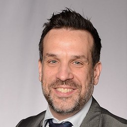
Dr. Igor Saftic, M.D., FEBTS
Clinical Director of Thoracic Surgery, University Hospitals Bristol and Weston NHS Foundation Trust Bristol, UK
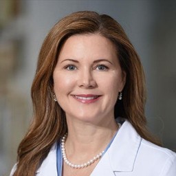
Prof. Shanda H. Blackmon, M.D., M.P.H., F.A.C.S.
Executive Director at Baylor College of Medicine Lung Institute, Houston, USA
About your hosts
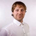
Pieter Slagmolen
Innovation Manager, Materialise

Sebastian De Boodt
Market Director, Materialise
Share on:
L-104584