
CUSTOMER STORY
How University Medical Center Göttingen Optimizes CMF Surgical Outcomes through Collaborative Case Planning
Transferring virtual surgical plans into the operating room as precisely as possible is a key challenge in CMF surgery, especially when information and responsibilities are shared between different teams. At University Medical Center Göttingen, this challenge has long been addressed through close interdisciplinary collaboration between the departments of Orthodontics and Oral & Maxillofacial Surgery (OMFS). Today, digital planning with Mimics Enlight CMF forms a central pillar of this collaboration.
Complex starting points require personalized solutions
In orthodontics, digital planning is primarily used for combined orthodontic–orthognathic treatments. These therapies are indicated when pronounced skeletal discrepancies between the maxilla and mandible can no longer be compensated by dental camouflage alone after completion of growth.
“Discrepancies may occur in the sagittal, transverse, and vertical dimensions — and in complex cases, in all three dimensions combined,” explains Dr. Johanna Trautmann, Specialist Orthodontist and Senior Physician at University Medical Center Göttingen. “The goal is always to achieve an optimal functional and aesthetic outcome on both the dental and skeletal levels.”
In oral and maxillofacial surgery, the focus is often on reconstruction of bony defects caused by oncologic resections, trauma, or inflammatory disease. Prof. Dr. Dr. Phillipp Brockmeyer, MBA, FEBOMFS, Senior Consultant and Deputy Medical Director of the Department of Oral and Maxillofacial Surgery, is responsible for reconstructive head and neck surgery. Planning is an integral part of that role.
“These defects require highly patient-specific planning,” he explained. “One of the key challenges is translating the virtual plan into the operating room as precisely as possible.”
“Each starting situation is highly individual and requires intensive interdisciplinary coordination. Digital planning tools help us visualize this complexity and make well-founded treatment decisions together.”
High demands on planning reliability and predictability
What unites both disciplines is the need for maximum planning reliability and for predictable outcomes. Beyond achieving stable occlusion, numerous additional factors must be taken into account: biologically feasible movement distances, relapse prevention, muscular and functional considerations, and increasingly, the airway situation, especially in patients with conditions such as obstructive sleep apnea.
“Each starting situation is highly individual and requires intensive interdisciplinary coordination,” says Dr. Trautmann. “Digital planning tools help us visualize this complexity and make well founded treatment decisions together.”
From established concepts to the next generation of digital planning
University Medical Center Göttingen has a long tradition in orthognathic planning. As early as the 1990s, they developed splint concepts to reliably transfer planned jaw positions and clinically registered condylar positions into the surgical setting. With ongoing digitalization, these concepts were continuously refined and eventually fully digitalized.
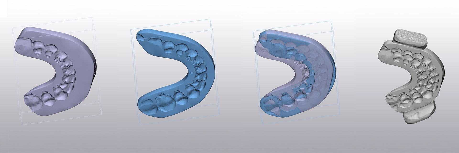
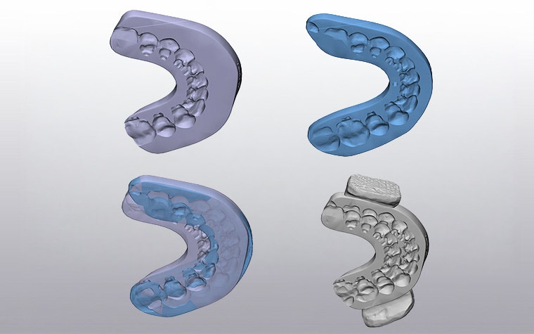
In Göttingen, the teams routinely apply a well-established concept for intraoperative condylar positioning. During surgery, they temporarily stabilize the condyle-bearing segment of the mandible using a special splint design featuring so-called “balconies.” This double impression splint captures both the preoperatively registered and the final planned mandibular position, allowing the condyle bearing segment to be fixed temporarily via the “balcony” during repositioning.
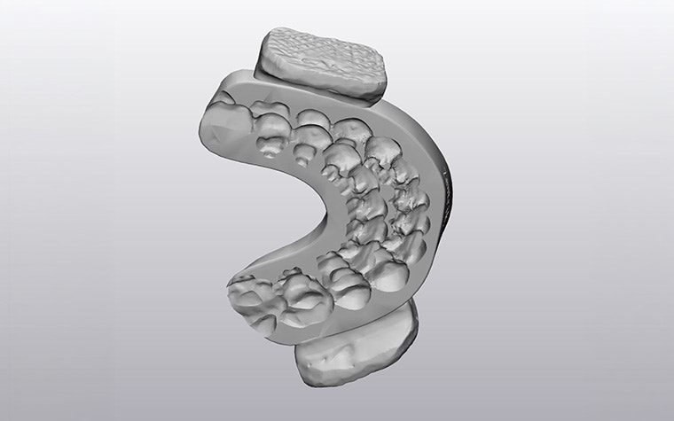
Such customized splint designs are not typically foreseen in conventional, highly workflow-driven software solutions. However, through close collaboration with Materialise, the teams can design these specialized splints in Materialise software, such as Mimics Enlight CMF and 3-matic.
Efficiency gains and new flexibility in daily clinical practice
One of the key advantages of Mimics Enlight CMF is the substantial increase in efficiency when creating anatomical 3D models. Many preparatory steps — such as segmenting key anatomical structures like teeth and the airway — are now automated during pre-planning.
“Previously, these steps took two to three hours. Today, we simply load scans and CBCT data into the software and start the workflow immediately,” explains Dr. Trautmann. “Overall, this results in time savings of approximately 70 percent.”
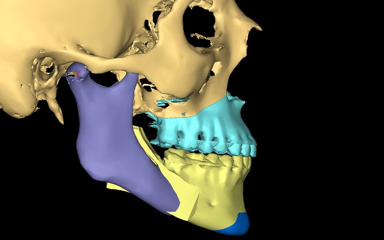
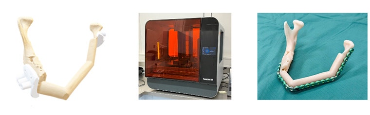
In CMF procedures, clinical teams can make virtual surgical plans for bony reconstructions quickly and in a standardized, validated manner. They can easily segment, virtually osteotomize, and semi-automatically position bone segments, plus export planning results as STL files and transfer them to 3 matic for the CAD design of osteotomy guides.
Improving accuracy and predictability in the operating room
Improved planning reliability has a direct impact on surgical execution. The high accuracy of splint fit means that intraoperative adjustments are rarely required, reducing chair time and improving the transfer of planned jaw movements into clinical reality.
In oral and maxillofacial surgery, digital planning in combination with CAD/CAM manufactured surgical guides contributes to improved predictability and execution of complex reconstructions.
“Automation reduces uncertainty and significantly increases safety, particularly in demanding cases,” says Prof. Brockmeyer.
Cross-team collaboration and improved communication
Another major advantage of digital planning is the shared visual basis for decision making. All planning steps are fully transparent and visualized in 3D, facilitating collaboration between orthodontists and surgeons and communication within the clinical team. This also supports patient education and involvement, with features such as soft-tissue simulations that make it easier to understand plans and expected results.
For clinical teams, this leads to simplified workflows combined with high precision. For patients, the main benefit lies in the accuracy of planning and execution — an essential prerequisite for stable functional and aesthetic long term outcomes.
Digital planning is an integral part of modern care
“Mimics Enlight CMF enables precise, efficient, and intuitive 3D planning that integrates seamlessly into established clinical workflows,” summarizes Dr. Trautmann.
From the CMF perspective, Prof. Brockmeyer adds:
“With Mimics Enlight CMF, we can efficiently plan even highly complex patient specific jaw reconstructions in a standardized and validated manner, without delaying the treatment pathway. We also see significant cost advantages compared to patient specific implants.”
Mimics Enlight CMF has become an integral part of daily clinical practice at University Medical Center Göttingen, enabling collaboration between departments and helping deliver better outcomes for patients.
L-105375-01
Share on:


You might also like
Never miss a story like this. Get curated content delivered straight to your inbox.