INTERVIEW
UMC Utrecht is on the Forefront of Innovation with Point-of-Care 3D Lab
Why does the University Medical Center (UMC) Utrecht in the Netherlands invest in the latest 3D technologies for their craniomaxillofacial (CMF) practice? To bridge the gap between research and the clinic and provide cutting-edge care by delivering 3D planning, 3D design of guides and models, technical support to surgeons, and technical information to patients all in one place.
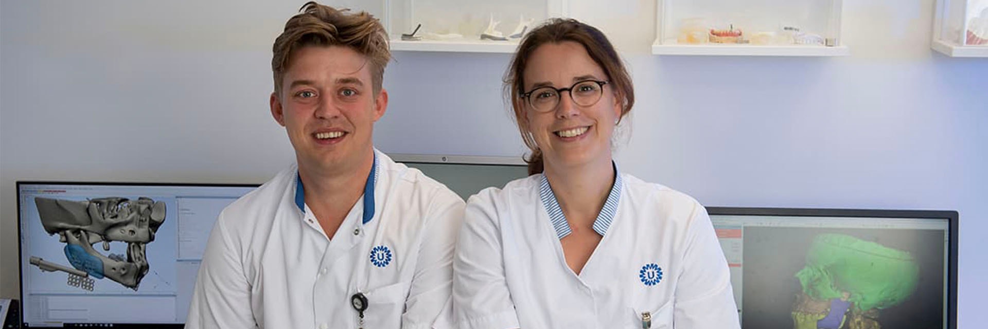
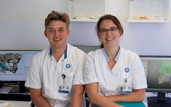
We recently sat down with Joël Kortes, and Maartje Kienhuis from UMC Utrecht’s 3D lab to hear how they went from using an analog dental lab until 2012, to their 3D FaceLab today.
What was the driver to change the in-house dental lab to a 3D technical lab?
The surgeons and dentists were the main drivers to change the analog dental lab into the 3D FaceLab. Most surgeons are hands-on people, but are not trained to use 3D computer software. They want to incorporate all the 3D possibilities but with the help of technicians. At first, a lot of the 3D work was outsourced. Now we have the in-house 3D service to make the process more convenient and to shorten lead times and reduce costs.
Today, our 3D FaceLab is the connection between the clinic and research as we have medical technicians from our lab working in the hospital. Because of this involvement, we are further able to understand how our research can be used in practice, and these findings are used to make our research even stronger.
Aside from time, money, and putting innovative research into clinical practice, the head of our department also sees the importance of 3D education. Our lab teaches doctors in training how to use 3D techniques to get the next generation of medical professionals working with the most innovative healthcare practices.
What are the other main advantages of having a 3D technical lab in-house?
Working in-house allows us to be involved in patient treatment, but also for monitoring the data of the patient. For example, to show the difference in dental models after the surgery but also use that data for the reconstruction and rehabilitation.
What we’ve seen is that the digital workflow benefits both the surgeons and the patients. Current and future patients not only want information about their illness and medical treatment plan, but also ask for insight into their technical treatment plan. A 3D visualization helps them to understand the technical part of their rehabilitation.
For the oncology patients, there is often great confidence in the medical procedure, but they remain uncertain about the aesthetic outcome. By involving the maxillofacial prosthetic (MFP) technician in the treatment plan and preparing a plan with the doctor, this information can be pre-operatively estimated and reduce this uncertainty.
Before the operation, 3D images can be used to show how the operation is planned and prepared digitally. Also, the figures and planning can be displayed for patient expectation management.
This way of working is also used for our orthognathic patients. They even tell us that an extensive conversation with a technician shows them the involvement of our entire team. We find that a lot of patients appreciate that part of our role in their treatment process.
How has this changed the way you work?
When we first started using these new technologies, the big concern was that sometimes your ideas were ahead of what the software could do. So I remember in the beginning we had a lot of ideas, but the computer said no. And now it’s fantastic that the only limitation is your individual know-how.
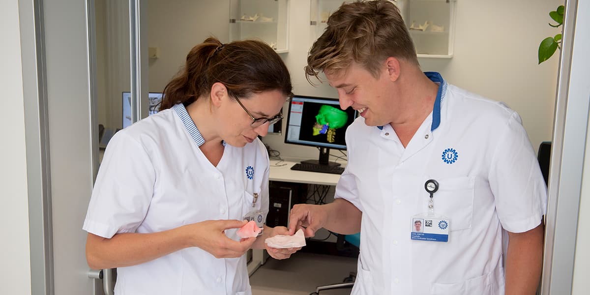
Today, before every 3D planning we meet the surgeon pre-operatively to discuss the position of the implants or the design of the guides. Once approved, we visualize the plan for the patient and surgeon and put the data in the patient file. When asked, we join the surgeon and patient in the clinic to explain the technical side of the intervention.
What exactly is the lab doing today?
Before 2012 our lab was a supportive analog dental lab for our CMF and special dental care department with a focus on oncologic patient care and the orthognathic surgery. Today, the MFP technicians do the entire 3D planning of our orthognathic operation and dental implants at our in-house 3D FaceLab and are more included in the patient care in the clinic.
Our daily activities are a combination of digital and analog. We use 3D techniques where it helps us to provide better patient care, but we use the analog ‘hands-on’ techniques when necessary. For example, the reparations of dentures or the fabrication of facial prosthesis from silicone are still mostly analog craftsman work.
Which software drives the operation of your lab?
Most of the time, we are using Materialise ProPlan CMF, Materialise 3-matic Medical and Materialise Mimics Medical. Also dental-specific software from 3Shape. With a combination of these software packages, we can design and manufacture our guides and models for patient care as well as for the simulation models the surgeons use.
The software helps us to work quickly and meet the short lead-time requirements. It’s easier to talk through the procedure with the dentist or the surgeon with this software. Because of our central location in the hospital, the doctors can easily access our lab.
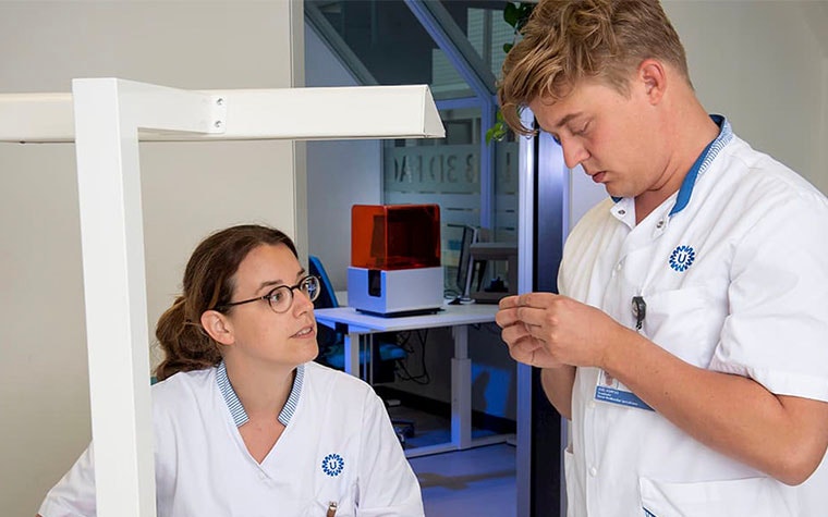
For the dental care department, we are now working with an intraoral scanner and software together with the use of qualified 3D printing materials.
Could you please give an example of a recent case?
We recently had a patient who had a partial resection of his maxilla. Because of our pre-operative planning, we could place a pre-designed surgical obturator, which are prosthetics used to fill openings in the mouth, such as missing teeth and soft tissue. And when he came for his first appointment we already had a new design and could place the new dental obturator with his teeth in their original location.
This is an optimization of our digital workflow; a consequence is that all models are digitally stored. This facilitates expeditious fabrication of a new obturator upon damage or need for adjustments. The data that we collect pre-operatively can be useful in further reconstruction, and the design of the 3D obturator can be adapted and re-manufactured.
Evaluation of the digital workflow will focus on optimization of the pre-operative design, reduction of discomfort for the patient, and cost-effectiveness.
Looking forward, what are the essential needs to support the continued growth of 3D printing in medicine?
We would like to take the lead to bring 3D towards other specialties in the hospital. Our lab is a well-organized starting point. Working with 3D specialists from collaborating departments can lead to innovation. Our main concern is that if we continue to scale up, we worry that we’ll lose contact with the patient, which is the key in our way of working today.
By acknowledging that contact with the patient on the technical part of the therapy is essential in providing good and effective care, we can really provide the best care we can. The personalized designs, or even the surgeon-specific designs, will only succeed when the MFP technician works in a 3D lab embedded in the clinic and close to the surgeons.
We’ve found that by using 3D services in our hospital that we’ve already made a difference. By staying with these technologies and moving them forward, we are committed to continue to do the best for our patients in the future.
Mimics Medical is not intended to predict the performance of a medical device.
Share on:
You might also like
Never miss a story like this. Get curated content delivered straight to your inbox.