CUSTOMER STORY
Anatomy Education at its Best: Printing and Painting Replicas of Donor Hearts

What do donor hearts and 3D Printing anatomy have in common? The answer to this is the University of Minnesota’s Visible Heart® Lab. Not content with simply teaching their students with 2D images, the team at the lab has moved their academic approach to a whole new level: 3D models of real human hearts. Imagine being able to train as a surgeon with a complete, tangible heart model, as opposed to learning off paper! And imagine acquiring the skills to make 3D models for any operation you might perform throughout your career? Here’s how Materialise enables the Visible Heart Lab’s unique approach to teaching, education and research.
Dr. Paul Iaizzo has headed the Visible Heart Lab at the University of Minnesota since 1990. The lab was created with the aim of educating students at the university, and driving research in the field of cardiology. From 1997 onwards, Dr. Iaizzo and his team began working on large mammalian isolated heart models in collaboration with Medtronic, and these days the laboratory is a unique place to perform translational systems physiology research, from cellular and tissue studies to organ and whole body investigations.
Anatomy education at the Visible Heart Lab
The Visible Heart Lab also works in collaboration with LifeSource, a non-profit organ donation organization from the Upper Midwest. When an organ donor’s heart is not suitable for transplant, the lab will receive the heart (with permission from the donor’s family) to use for research. If the heart is mostly intact (including the presence of the great vessels) and can perform a reasonable hemodynamic function, the researchers at the lab will reanimate it, using Visible Heart methodologies.
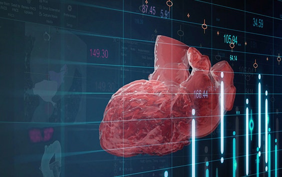
Each heart is subsequently preserved in an anatomical end-diastolic state, and used as an anatomical specimen. As there are no consequences involved in harming the heart of a dead patient, it is possible to submit it to a high dose of scans – micro-CT and MRIs are performed – which results in extremely high-quality, precise image scan sets. These images (in the form of DICOM files) are subsequently uploaded into Materialise Mimics software and transformed into an equally accurate 3D model which can then be printed. The lab has a veritable heart model library, stocked with everything from models of dilated cardiomyopathy to normal cardiac structures. A de-identified patient history is available for each specimen, and additional pre-recovery patient information is available in some cases, including the results of echocardiography and electrocardiograms.
The potential for 3D-printed models
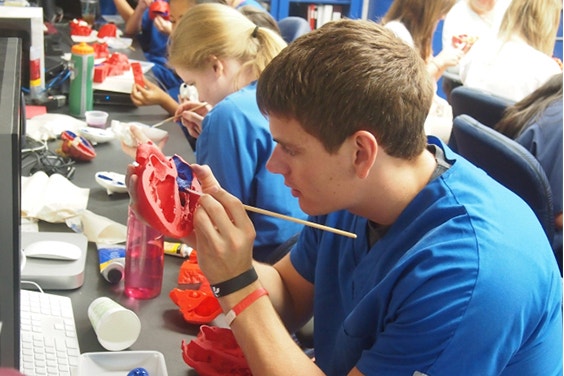
Dr. Iaizzo believes that these 3D-printed heart models are a great education tool, and the students at the lab are expected to learn the entire process, from using the 3D Printing software to painting the finished models. He believes that mastering these tools gives them an academic advantage, as they can get a complete overview of the organ and see first-hand how they would go about planning a medical device delivery or surgical procedure. And it doesn’t stop there: doctors can use this technology to scan a patient’s diseased heart before the operation, scan it again after the operation to see how the heart has been repaired and then further reverse remodel it.
A selection of these highly accurate 3D-printed models can now be ordered through our website, as part of a long-standing partnership between the University of Minnesota and Materialise.
Share on:
You might also like
Never miss a story like this. Get curated content delivered straight to your inbox.