EXPERT INSIGHT
Personalized Patient Care in Veterinary Neurology Becomes a Reality Thanks to 3D Printing

Pets are beloved family members, providing comfort, joy, and companionship to millions. In fact, around 88 million households owned a pet last year in the EU alone.1 So, when these precious animals need treatment, it can be very distressing for the patients and their owners, especially if surgery is needed. However, 3D printing techniques and software are changing the face of veterinary surgery. 3D printing offers personalized patient care that delivers more accurate and reliable outcomes for the patient and surgeon — while also facilitating better communication with pet owners about the procedures.
Dr. Julien Guevar, Veterinary Neurologist and Neurosurgeon at the University of Bern and AniCura, Switzerland, is an advocate of 3D printing in veterinary medicine. He regularly uses Materialise Mimics to help prepare for his surgical procedures more effectively. We spoke to him to learn about the benefits of 3D printing in the veterinary world, its untapped potential in certain regions, and how it has improved his preparation and surgical procedures.
Thanks for joining us, Julien. Could you tell us a bit about your background and research?
Well, I’m a veterinary neurologist and neurosurgeon who’s always been interested in innovation in spinal surgery. My goal is to identify ways of making surgeries safer and more accurate for my patients (mainly cats and dogs). And this research is in combination with veterinary orthopaedists and bioengineers.
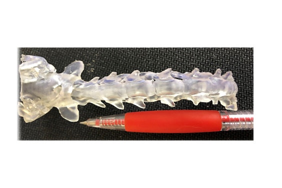
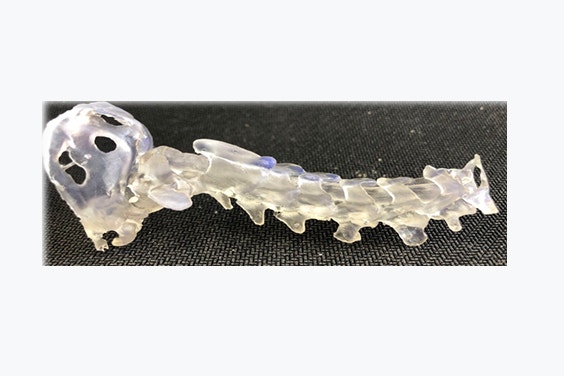
3D-printed model of the cervical spine and caudal part of the skull of a toy breed dog with atlanto-axial subluxation, with and without a pen to illustrate scale.
I’ve studied in Belgium and worked in Europe, the UK, and the USA; however, my interest in 3D printing was sparked in the US at North Carolina (NC) State University. At NC State, with the help of veterinary colleagues and engineers, I was able to see the power of interdisciplinary collaboration. Seeing such enthusiasm about the technology and the diversity of its applications really piqued my interest. My passion grew even further when I arrived at the University of Wisconsin-Madison, as they were deeply invested in 3D printing at the time and were long-term users of Materialise’s software. This was also the place where I had the opportunity to be part of a small 3D printing lab on campus.
3D printing is becoming increasingly popular in the veterinary sector. Could you tell us what made you use this technology and describe the major benefits for veterinarians?
3D printing is very accurate, allowing me to prepare for my surgery more safely and efficiently. For example, I can plan and prepare my surgeries in advance, anticipate where and how the implants will be placed, and even create drill guides with specific trajectories. Investing a little time before surgery saves me a lot of time during surgery, as my thought process was conducted beforehand. This type of knowledge and awareness makes the procedure more predictable — as I visualize exactly where and how I need to operate — and therefore safer for the patient. In comparison, the technique we have mostly used when placing implants into the vertebrae of dogs up to now is eyeballing the starting point and the angle for drilling, based on data from the CT. We can do better than that.
However, when using Materialise’s software, I can efficiently prepare my surgeries, aiming for a safe and optimal trajectory. This is particularly important when using drill guides and operating on very small patients. Sometimes, these animals weigh less than 2 pounds, with spinal cords that have widths smaller than a pencil. These bones are smaller than any of the ones you would find in human pediatric surgery, so precision is crucial.
Also, as communication with the pet owners and their understanding of these complex surgeries is paramount, veterinary surgeons can physically show and explain to the owner what will happen during the procedure with the 3D-printed models. For instance, indicating to the owner that a screw placed two millimeters in the wrong place could damage the artery or the spinal cord, respectively, reveals why precision matters — it’s not hard to convince them after that. 3D printing increases a surgeon’s margin of safety and allows us to provide patient-centric care that is almost comparable to human medicine. Not to mention, 3D printing is a financially viable solution nowadays.
For me, 3D printing ticks all the boxes: it’s extremely safe, reduces our time in the OR, and improves accuracy — leading to better patient outcomes.
What are the main differences between the US and Europe in terms of personalized tools for veterinarians, and can you give us some reasons for the differences?
In my experience, the advancement of veterinary surgery is similar wherever I’ve been. But the biggest difference is in culture. This is not the case for everybody, but in general, in the UK and USA, they see the potential and they go for it. In Europe, however, we first see how complex it will be to reach that goal and how it might disrupt old habits. It’s all in the mindset. The positive thing is that the wind is turning, and some pioneering colleagues from the universities of Utrecht and Zurich have been tapping into that potential. We now need to increase the awareness of the benefits of this technology outside of the academic realm. In the UK and US, owners have heard about the benefits of this technology through the media and demand the use of 3D printing, whereas, on the old continent, awareness is still rather limited. We need to work on that; it’s in our patients’ interest.
How did Mimics software support you during your work/surgical planning?
Mimics was the go-to software for my former colleagues in America, and that’s where I first learned about Materialise. Now, it’s the default option for me. I have the chance to work in academia and the private sector, and 3D printing has a big role to play in both places.
But, before embarking on live surgeries and claiming a technology to be safe and accurate, as an academic and clinician, I had first to demonstrate those facts and expose them to the scientific community. I cannot advocate for something I have not tested. While researching the benefits of personalized 3D-printed drill guides, I had the chance to collaborate with an application specialist at Materialise who assisted me with the most appropriate and accurate way to use the software. It was absolutely wonderful; we reviewed the drill guide workflow and optimized it. Now, it takes me 15 minutes from processing the CT data to hitting the print button of my Formlab printer. This was, for me, an essential step in developing better surgeries for my patients, and it must be done with quality control. So, practically speaking, if I see a patient in need of drill guides for surgery, I can design and print surgical models overnight and be ready for surgery the following morning.
In my view, if I have a guide that can tell me exactly how and where to place an implant because I’ve done all the preparation beforehand, then I’m going into the OR with the right tool.
Could you go through your process step by step?
Essentially, my process is split into the preparatory design, pre-op testing, and surgical phases. The latter is shorter because all the thinking was done prior. Let’s take two examples. With the CT data of a patient with a vertebral column problem, I use Mimics Core to create a 3D model of the vertebra and establish my safe trajectories. I export this data into 3-matic, where I can design the drill guides. I then usually print a set of vertebrae and drill guides in clear resin using a Formlab printer and assess their fit or any printing error as if I were already in surgery. There are usually no surprises, so then I print them in a biocompatible resin that can be used intraoperatively and that I know won’t be altered during the sterilization process. I can also take a 3D-printed model in the OR with me so that I can see clearly while physically holding the vertebrae in front of my eyes.
It can also be used for other surgeries, of course. If I need to remove a brain tumor in a dog or a cat. I could print the skull and the tumor for use in surgery, allowing me to precisely identify important anatomical landmarks on the model and the patient, to reach and remove them more efficiently. The same was true for dogs with very large skull tumors that needed rather extensive craniectomies. Little things like that can help make a major surgery easier and a lot safer.
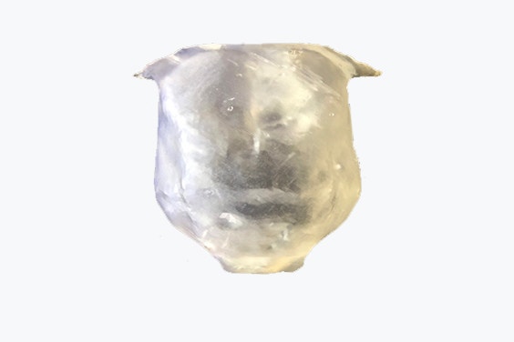
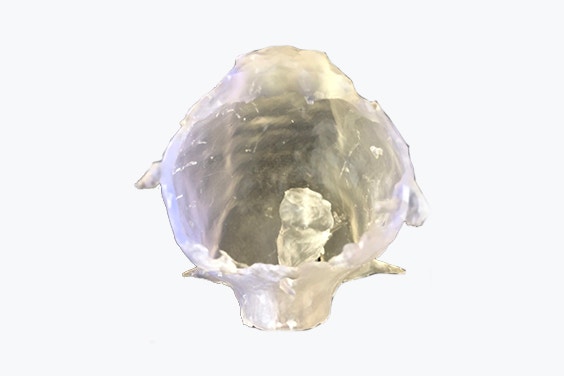
3D-printed model of the skull of a dog with a brain tumor.
What is your next goal/research topic?
There are two aspects to this question. The first one, which we have already discussed, is the quest for safer and more individualized surgical solutions. We should continue researching new solutions for old problems. And this is not only for small animals. Colleagues who treat larger animals could surely benefit from this in their daily surgical practice, too. Having brainstormed with some of them, we know the need exists and solutions can be designed. The research published in veterinary medicine in the 3D printing field is currently small, yet the findings are unanimous; it is undoubtedly powerful for accurate and safe surgery. For example, research papers at NC State proved that students reached the same level of accuracy and safety as trained neurosurgeons when using a drill guide for vertebral column surgeries.2, 3 This quest is no different from our human peers. We all want accurate and safe techniques for our patients. The gold standard in human neurosurgery is computer-assisted neuronavigation, and soon robotic-assisted neurosurgery. Although they are fantastic technologies, they are also cost-prohibitive for most veterinary practices. This, in my opinion, is another box that 3D printing ticks. It is affordable.
The second one, which I am also very fond of, is the use of these models for education. There is an added value to 3D printing in academia, as the models could be used by residents in training to practice with and prepare for complex and/or uncommon surgeries. Normally, there’s a steep learning curve, but 3D printing can reduce this significantly. The big advantage is that residents can visualize in 3D and touch a model instead of relying on a textbook. It won't be long until we start printing vertebral columns, muscle, and skin models to practice and train on.
Overall, the advantages of embracing 3D printing are very clear. I see many benefits, such as improving pupils’ education, pet owners’ awareness, the surgical training carried out, and last but not least, the safety of neurosurgery for cats and dogs. Innovation to improve animal health is my professional passion.
[1] The European pet food industry (FEDIAF) facts and figures 2020
[2] Accuracy and safety of three-dimensionally printed animal-specific drill guides for thoracolumbar vertebral column instrumentation in dogs: Bilateral and unilateral designs
[3] Accuracy of three-dimensionally printed animal-specific drill guides for implant placement in canine thoracic vertebrae: A cadaveric study
L-102442
Share on:
You might also like
Never miss a story like this. Get curated content delivered straight to your inbox.