PATIENT STORY
How a 3D Lab Improves Surgical Outcomes in Top Pediatric Hospital
At Rome’s Bambino Gesù pediatric hospital, the 3D lab has grown into a formidable support for the surgical teams. Anatomical 3D reconstructions give them a better view on each patient’s case, allowing them to discuss and prepare cases beforehand. This shortens the actual surgery and improves patient outcomes. We spoke with Dr. Aurelio Secinaro and Luca Borro, two senior experts at the hospital. They testify to the many advantages of 3D reconstructions with Materialise’s Mimics Innovation Suite, software that helps with even the most complex of surgeries. They explained how it was instrumental in the successful separation of twins with conjoined brains.
The ‘Ospedale Pediatrico Bambino Gesù’ is reputed for its pediatric expertise. It offers more than 20 specialties through 10 pediatric departments and treats children not only from Rome or Italy but even from neighboring European countries. In addition, its humanitarian program serves people in 16 countries on four continents.
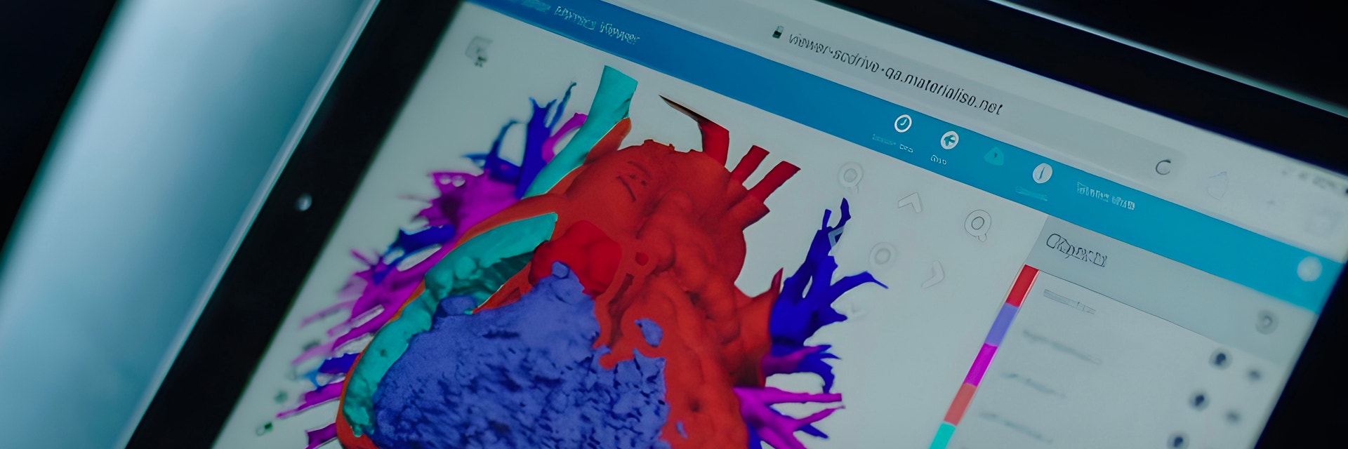
Luca Borro is a Biomedical Engineer at Bambino Gesù. He’s helping to build and run its 3D lab and therefore is a firsthand witness to the many benefits of 3D reconstruction. “In our hospital,” he says, “we have a dedicated 3D lab with a reconstruction workstation running Materialise’s Mimics Innovation Suite and a graphics tablet with which we segment complex clinical cases. We also have two FDM 3D printers that we use to print anatomical models that need to be produced urgently.
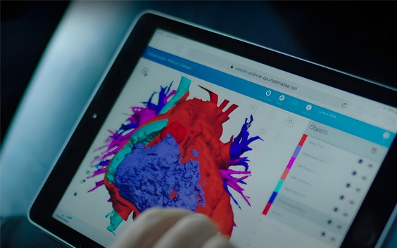
“The 3D lab is fully engaged with 3D reconstructions of anatomical parts based on radiological images, reconstructions that are requested by the hospital’s various departments. Mimics software allows us to perform high-quality complex segmentations quickly. Project management with Mimics is super smooth, and thanks to the Mimics Web Viewer we can quickly and easily share 3D models with everyone on the surgical teams.”
Better surgery
Luca’s colleague Dr. Aurelio Secinaro, director of Bambino Gesù’s cardio-thoracic imaging unit, welcomes the new techniques as no less than a revolution.
“I am a specialist in diagnostic imaging,” says Dr. Secinaro. “I’m passionate and dedicated to the use of advanced techniques such as computed tomography (CT) and magnetic resonance imaging (MRI) in the diagnosis of heart, lungs, and other chest organs. Adding 3D techniques on top has revolutionized the field, as we can now study patients’ anatomy and pathology in three dimensions, with added depth and volume.
“The greatest advantage of using 3D models, whether virtual or printed, is that they help prepare for surgery. They allow us to make a better clinical interpretation of the problem and we use them to discuss and exchange views among the team members. As a result, the team eventually enters the operating room with more confidence. And the more complex a case is, the more this advantage plays out.
“In addition, the 3D models help to reduce the time spent in surgery. In the case of maxillofacial procedures, for example, we operate about 40% faster. That has immediate and tangible benefits such as a decrease in time under anesthesia and a lower probability of bleeding. In short: a better surgical outcome.”
Top clinical use cases
Bambino Gesù deploys 3D techniques mainly for maxillofacial surgery (mouth, jaws, face, and neck), heart surgery, and neurosurgery. “These are the medical domains,” says Dr. Secinaro, “in which 3D technology is clinically most useful and brings the greatest advantages.
“For maxillofacial surgery, we create and use 3D models to prepare for highly individualized, reconstructive surgery, for example, to repair craniofacial bone growth disorders or orthognathic disorders such as facial asymmetries and jaw retraction or advancement. In addition, we frequently do 3D reconstructions to prepare for facial bone reconstruction after accidents, or for cancer surgery. In maxillofacial surgery, we always print the models created with Mimics.”
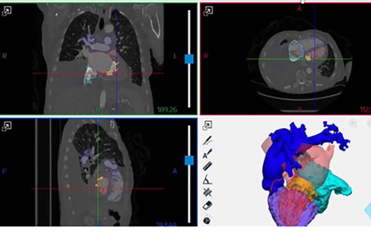
In cardiac surgery, the hospital’s experts use 3D reconstruction to get a better view of complex congenital heart issues in children and adolescents, says Dr. Secinaro. “These are for example tetralogies of Fallot, double exit right ventricles, transposition of the great arteries, or pulmonary atresias with interventricular defects. Except for very special cases, we create the models with Mimics but do not print them: the medical teams study the cases on the virtual models.
“Also, in neurosurgery, 3D reconstruction plays an essential role. Here we often see patients with very complex issues, requiring long preparation and a multidisciplinary team. An example was the successful separation of conjoined twins, where we needed to combine 3D technology with our top medical expertise.”
Conjoined twins — a most complex case
Ervina and Prefina, twin sisters, were born in the summer of 2018 in a village close to Bangui, the capital of the Central African Republic. Sadly, they were joined together at the head with what experts call one of the rarest and most complex forms of cranial and cerebral fusion, a total posterior craniopagus.
By a stroke of luck, Mariella Enoc, president of Rome’s pediatric Bambino Gesù Hospital, met with the twins and their mother Ermine in Bangui shortly after the birth. She decided to bring the girls to Italy for surgery at Bambino Gesù.
A multidisciplinary team of neurosurgeons, anesthesiologists, engineers, and plastic surgeons then started studying and preparing to separate the twins. The real challenge for the doctors was not so much that their skulls were joined, as that they also shared brain membranes and a significant portion of their brains’ venous system.
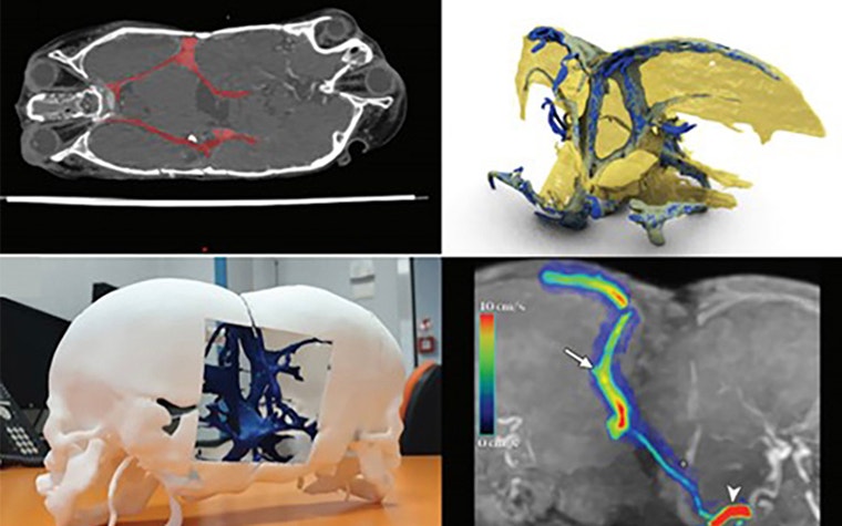
A highly complex and unusual model
Reconstructing a 3D model of the conjoined twins proved highly complex. There were numerous anatomical parts that had to be modeled. In addition, these were arranged in unusual positions, positions never seen before and so unique to this case of conjoined brains.
According to Luca, Mimics software was instrumental in making the 3D reconstruction process a success. It allowed for optimized workflows and manageable 3D mesh surfaces that were easy to work with. Also essential were the numerous editing functions and masks for segmentation, such as Boolean functions, split functions, and 3D segmentation functions. In a very real sense, these allowed the team to achieve an initial virtual separation of the twins’ brain and vein systems.
“The Mimics software,” concludes Luca, “optimized both the quality of work and the time it took to obtain the final model.”
With the model, the team could start laying out the step-by-step procedure for the separation. Also, most importantly, they could show the twins’ mother how the procedure would unfold, not on 2D images, but on a life-sized composite model.
Eventually, the complete separation was successfully realized, in a procedure that took 18 hours and involved a team of 30 people. According to the hospital, the two-year-old sisters were expected to make a full recovery.
The outlook for personal care through 3D
The use of 3D technology has seen a considerable acceleration in the last 10 years. According to Luca, that is because the recognition of the clinical benefits for patients and doctors has kept on growing.
“The accuracy and immediate clinical utility of this technology have helped spread it around the world. Every advanced hospital today uses complex 3D reconstructions and 3D printing for the clinical management of its patients. But there is more to come: virtual 3D reconstruction, for example, is transitioning from on-screen visualization of 3D anatomy to the immersive reality of viewers and holograms. All of this will help make 3D even more readable for the physician and provide considerable technological support for important clinical decisions.”
In a broader perspective, Dr. Secinaro sees 3D technology evolving into a high-tech point of care available in every children's hospital to serve their wards and clinical needs. “There will be 3D printers on every ward, where staff pick up patients' anatomical models and parts. Each hospital will likely also have virtual 3D stations where 3D reconstructions are created with software such as Materialise’s Mimics Innovation Suite. Reconstructions that can be managed in real-time and discussed remotely with other clinical centers in a fully immersive environment.”
L-102516-01
Share on:
You might also like
Never miss a story like this. Get curated content delivered straight to your inbox.