Enhance your segmentation workflow
Save time
Reduce manual effort and accelerate case processing by identifying and categorizing relevant information with AI.
Easily manage clinical case inflow
Deliver accurate 3D models more confidently and without sacrificing quality, especially in caseload peaks.
Optimize your team’s expertise
Spend less time on segmentation and more time on high-value tasks — improving consistency and reducing training time.
Get more out of Mimics with AI
Seamless connection to a complete platform
Automated segmentation is just a few clicks away from your workspace with integrated algorithms in Mimics.
Suitable for clinical use
Use AI-enabled segmentation with confidence, knowing it’s been cleared for medical use.
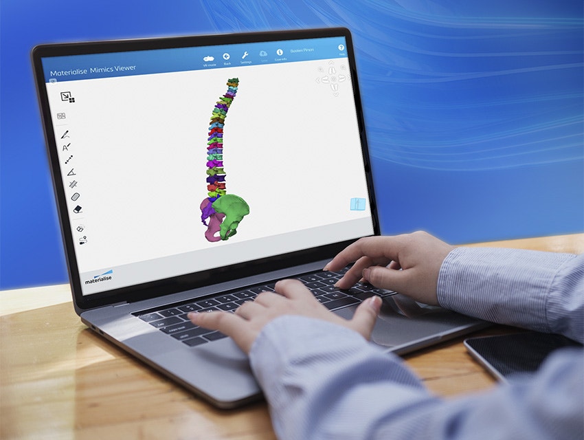
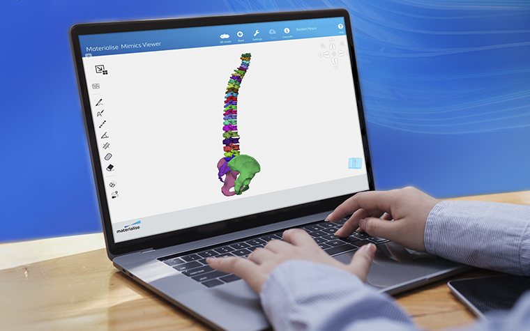
Smoother collaboration
Easily share the results with your colleagues to review the case, leave annotations, perform measurements, and more — they don’t even need a Mimics license.
Maintain control
After automated segmentation, you remain in control of your model. Use Mimics’ editing tools to fine-tune any results that don’t fully meet your needs.
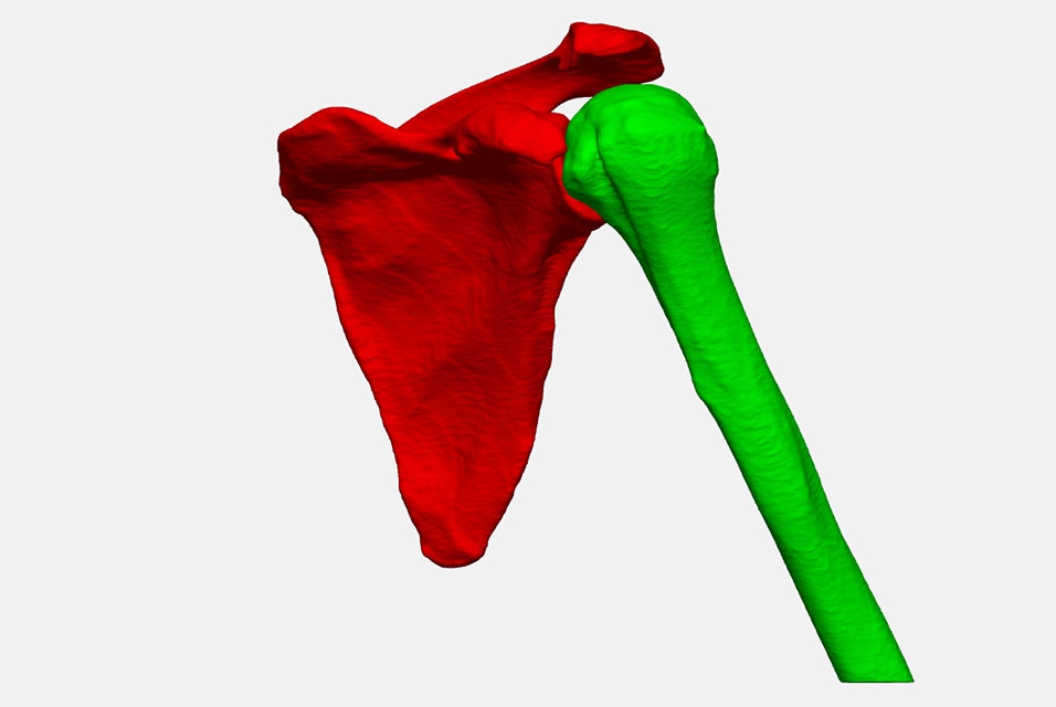
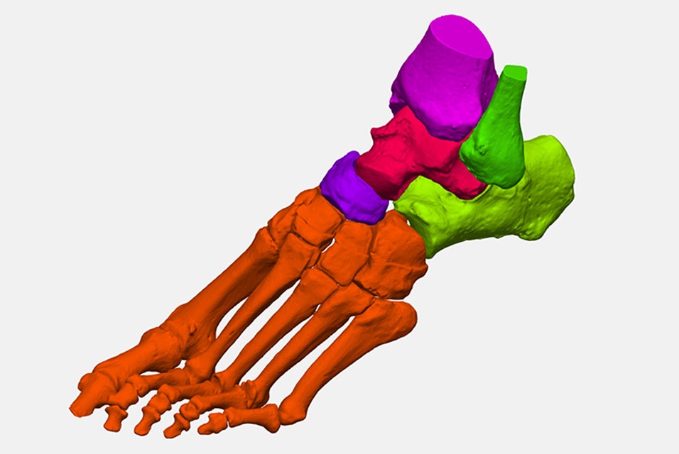
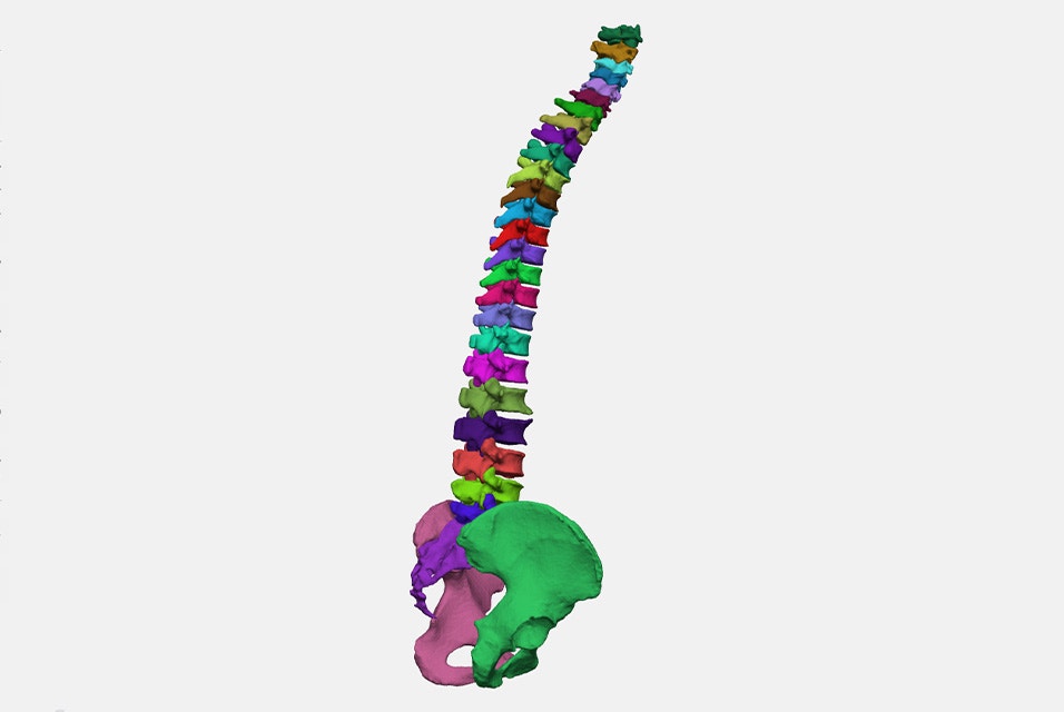
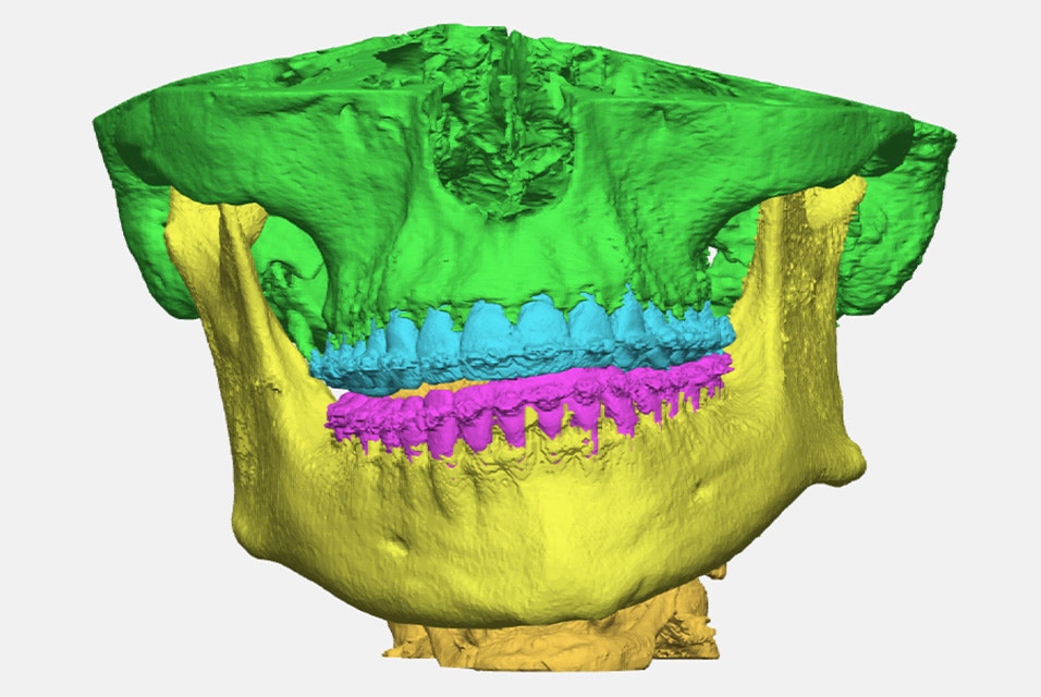
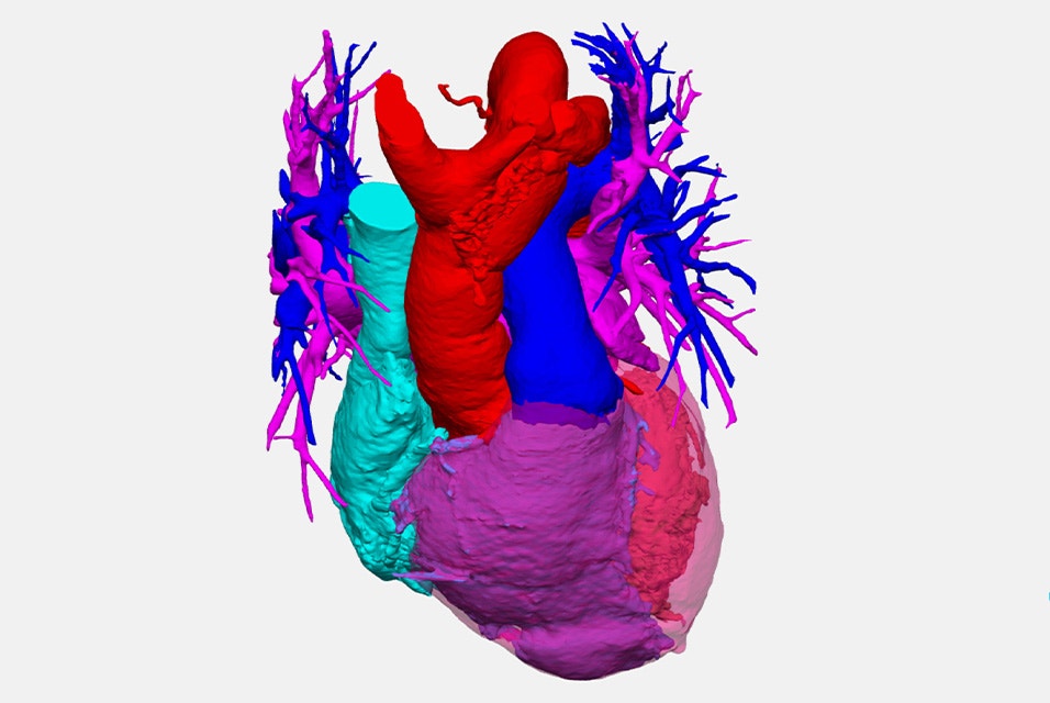
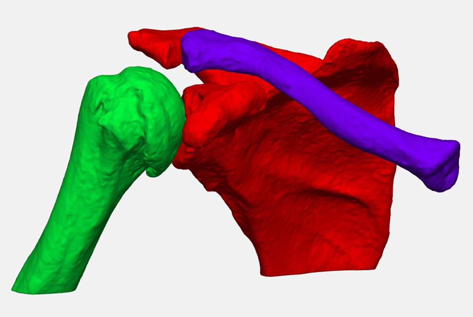
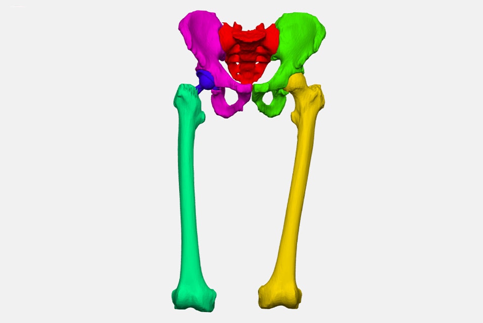
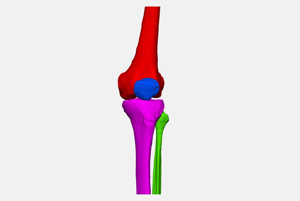
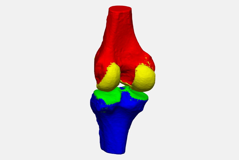
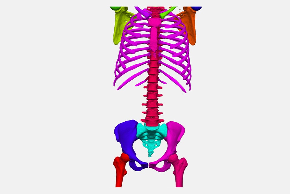
Available algorithms
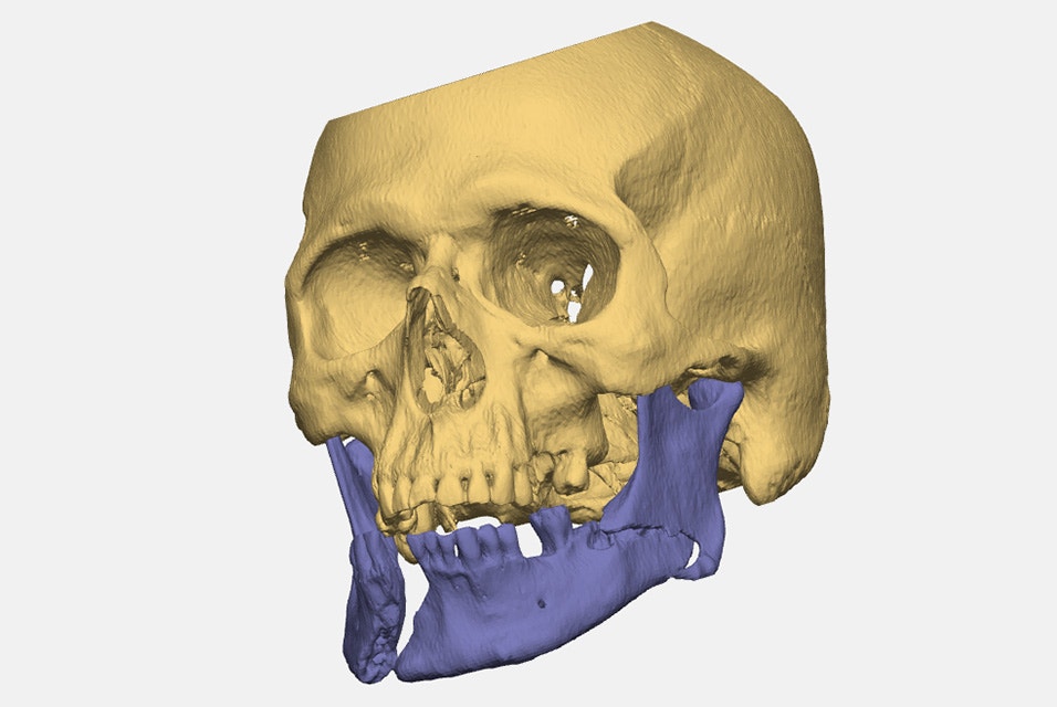
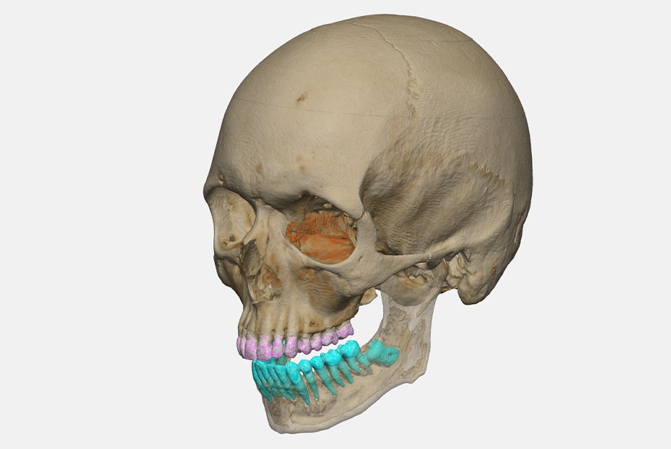
Where AI-enabled segmentation makes an impact
Go beyond visualization and interact directly with the anatomy to bring greater clarity to in-house preoperative planning.
Access industry-leading tools for biomedical research and education.
Build and scale your personalized medical device business with tools that support you from R&D to case management.
Scale new therapies faster and more effectively with AI-driven 3D insights for accurate segmentation and anatomical measurements.
Get inspired
L-104099-02
Please note that ‘AI-enabled’ refers to numerical algorithms using artificial intelligence, including thresholding, machine learning, deep learning, graph-based and model-based segmentation algorithms, and combinations thereof that have been locked and validated before release. It does not refer to any form of adaptive AI/ML.
Materialise Mimics mentioned above is a research version only. Not all the Mimics Automatic Algorithms may be available in your area for medical use.
Materialise medical device software may not be available in all markets because product availability is subject to the regulatory and/or medical practices in individual markets. In countries where no regulatory registration is obtained for Materialise Mimics and/or Mimics Viewer, a research version is available.
Please contact your Materialise representative if you have questions about the availability of Materialise medical device software in your area. For more detailed technical information, please refer to the technical documentation available in Mimics Viewer’s Help section.
Related products and services
Efficiently and accurately segment 3D medical images for virtual planning and analysis.
Easily view and share 3D plans, device designs, and anatomical models in this online platform.
Experience effortless online storage and case sharing with this cloud-based collaboration platform.