CASE STUDY
Total Artificial Heart Implantation Planning
The utilization of total artificial hearts in pediatric cases is limited by strict recommendations on anatomical characteristics for proper fit. The Phoenix Children’s Hospital uses Materialise Mimics to screen pediatric patients who fall outside the recommended range.

Industry
Healthcare
Key solutions
Materialise Mimics
The impact
Virtual fit analysis
Mimics is increasing the number of patients who can benefit from bridge-to-transplant (temporary total artificial heart) devices through patient-specific virtual fit analysis. Mechanical Circulatory Support (MCS) has been used in pediatric patients since the late 1980s as a bridge to heart transplantation, yet the utilization of total artificial hearts (TAH) or ventricular assist devices (VAD) in pediatric cases is still limited by strict recommendations on anatomical characteristics for proper fit. The Phoenix Children’s Hospital has identified a method of screening pediatric patients by using Mimics to determine if SynCardia’s temporary total artificial heart (TAH-t) may provide a solution for those who fall outside the recommended range.
In need of a new heart
A 14-year-old patient was admitted to the Phoenix Children’s Hospital with dilated cardiomyopathy, severe biventricular dysfunction, and ventricular arrhythmias. As no donor was immediately available, the teenager was placed on the heart transplant list and considered for a temporary total artificial heart.
Normally, the recommended minimum body surface area for the proper fit and function of the TAH-t is 1.7 m2 and 10 cm for the anteroposterior distance between the sternum and the spine at the T10 level. Though this patient was beneath the recommended fit criteria for the 70 cc SynCardia TAH-t, the clinical team was still able to proceed with the necessary TAH-t implantation based on their assessment of the preoperative CT scan data.
Implanting the SynCardia TAH-t
A sternotomy was performed on the patient, the ventricles and all four valves were excised, and the TAH-t was implanted successfully.
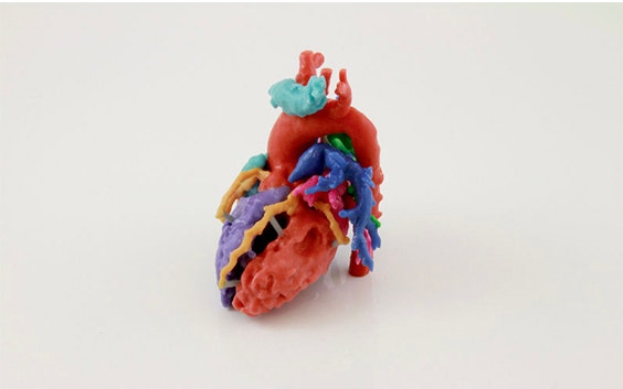
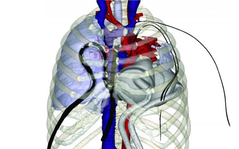
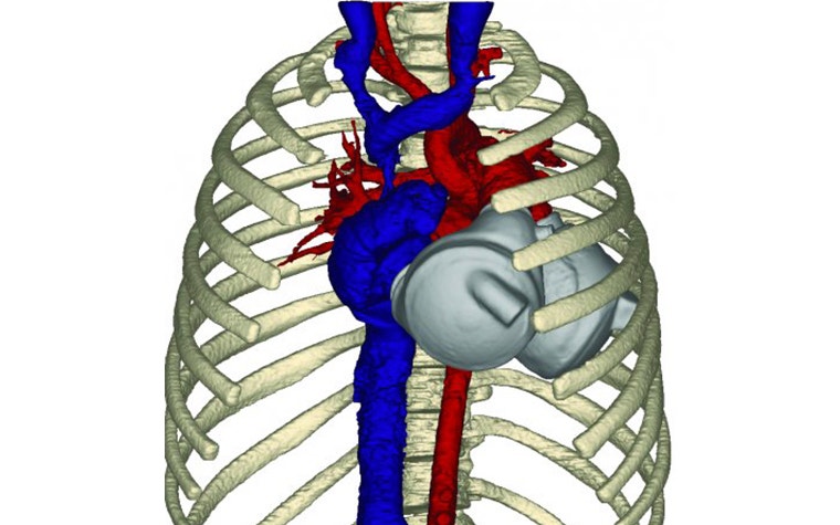
Postoperatively, complications arose. A post-implantation chest radiograph revealed left lung opacification which was confirmed by a contrast-enhanced CT of the thoracic cavity. In an effort to analyze possible obstruction from the TAH-t of pulmonary vasculature and bronchi, a 3D reconstruction was generated using Mimics. The cardiovascular, respiratory, and skeletal structures were all segmented in a 3D model. In addition, the blood volume was segmented from within the TAH-t device (the only portion visible on the CT scan). To obtain a complete model of the TAH-t, the device was scanned with a laser scanner. The surface mesh was then brought into Mimics and aligned to the blood volume of the device. This confirmed that the size and location of the TAH-t were not constricting any major cardiovascular or respiratory structures.
The role of the mimics in virtual fit analysis
A 3D reconstruction from a chest CT was crucial for visualizing the pulmonary arteries and veins as well as the bronchial structures to rule out compression from a TAH-t. This process showed that virtual modeling can assist with root cause analysis of acute complications that pediatric patients are experiencing. During this case, they also determined that virtual modeling of preoperative CT data in Mimics helps predict the appropriate fit of the device for future interventions. Pre-implantation fit analysis has already been applied in multiple pediatric cases where patients were excluded based on general fit criteria. These successes were made possible by utilizing CT and 3D modeling for a virtual or device fit analysis.
Mimics has made the virtual fit analysis process easy. Clinical teams simply create a 3D reconstruction of the patient’s anatomy from a preoperative CT scan. A laser scan of the SynCardia TAH-t (or other TAH/VAD device) is then brought into Mimics and positioned in place of the virtually excised organ. Based on a series of measurements and clinical review, the team determines whether a total artificial heart would properly fit within the chest cavity.
This process is opening up possibilities for pediatric populations who have few clinical options.
D. Bradford Sanders, chief perfusionist at Phoenix Children’s Hospital expressed, “CAT-scan imaging of the chest, in conjunction with Mimics, helped to determine whether the SynCardia Total Artificial Heart could actually be placed in the child’s chest.”
Share on: