CUSTOMER STORY
DJO Optimizes Hip Implant with Image-Based Population Analysis
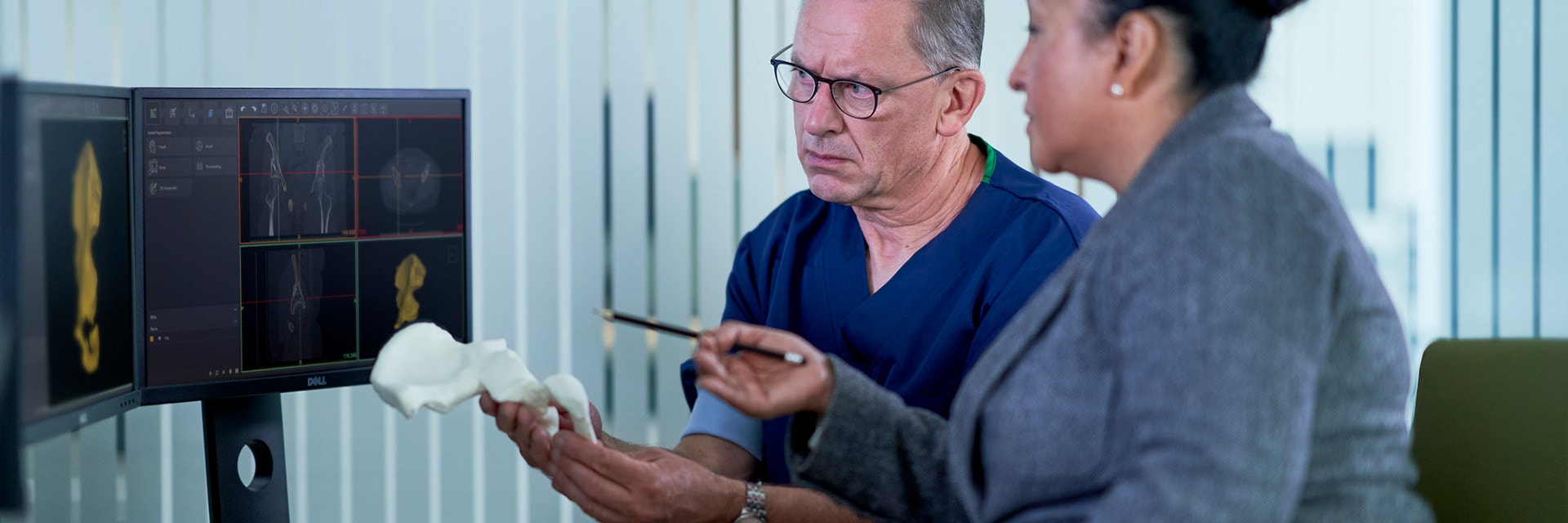
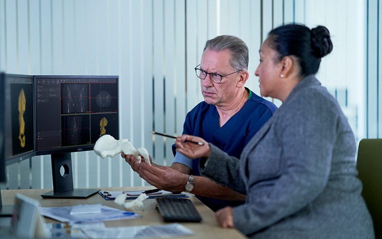
American surgical device company DJO was having challenges prior to the launch of their new implant, the TaperFill™ Hip Stem, a shorter femur stem designed to be inserted through a direct anterior approach, hereby sparing the critical posterior soft tissue. The design of the implant proved to be very tricky as it needed to fit closely in the cortical bone to ensure stability. As there was not much room for error, it was difficult to create a design that fit a maximum amount of patients, since anatomy differs slightly from person to person. DJO optimized the hip implant with the help of image-based population analysis.
DJO has a long history of experience with implants, but traditional anatomic information (literature reports, measurements on X-ray images, cadaver studies) was proving to be not enough in this case. Femoral fractures were reported during the implantation of the new device in the first few cadaver labs. As the market release deadline of the product rapidly approached, each member of the engineering team drew on their own individual experience, leading to different possible root causes for the fractures.
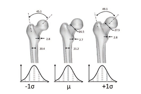
In order to have a more objective approach in resolving the design issue, DJO turned to Materialise. To help DJO answer that question, Materialise’s ADAM services team performed a population analysis study. They started from a database of femur CT scans. A library of virtual 3D femur models of the patient population was created. Using their expertise in statistical shape modelling, the average femur shape and typical smaller and larger femur shapes were created. In doing so, Materialise was able to capture the main shape variation present in a large database of scans, in just a handful of 3D femur models that were provided to DJO.
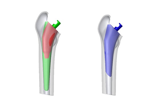
The engineers at DJO then virtually implanted their design into the population shape models. With this fit analysis they discovered that the design was too wide at a certain location along the stem, leading to local stress concentrations in the femur.
Using these population models and the results of this analysis, DJO made critical adjustments to their design. This new design has been a resounding success after 5,400 implantations. Since this case, the engineers at DJO have been using Materialise ADAM services for 3D population-driven design earlier in the design process. This is giving them an edge in anatomical insight and bringing innovative new designs to the market faster.
“Now that we have experienced the power of this technology firsthand, it is like we have a hammer and everything becomes a nail,” says Bryan Kirking, Surgical Biomechanics and Testing Scientist at DJO. “We have used this technology to improve projects throughout our R&D department at DJO.”
This story is just one example of how medical image-based population analysis is transforming the way medical device companies are developing their standard implant today. By having a virtual representation of the patient population and the way to extract the right design inputs, development teams are gaining a much deeper insight in anatomical variation. Now, they can safely try out new design concepts virtually before
L-102648-01
Share on:
You might also like
Never miss a story like this. Get curated content delivered straight to your inbox.