INTERVIEW
The Best Method to Capture Foot Anatomy? How Combining 2D and 3D Technology Help Create Better Orthotics
Orthotics have come a long way from being made with plaster casts and foam boxes. The process of making custom insoles is transforming as digital workflows bring more efficiency and accuracy to clinicians’ practices.
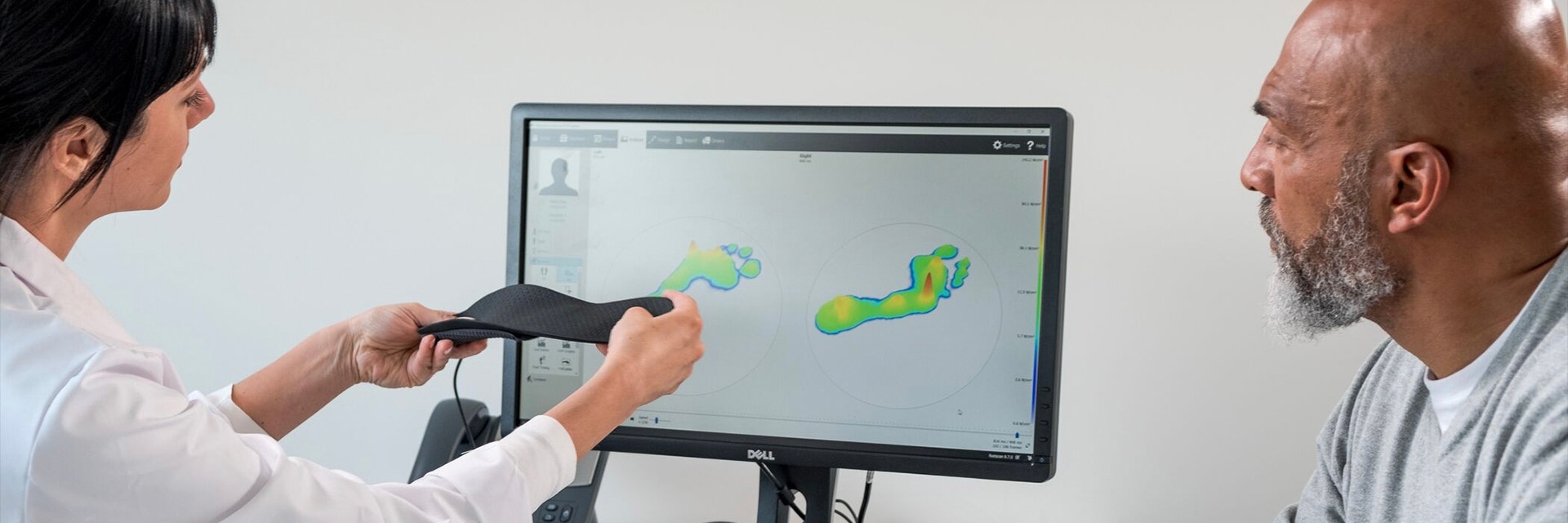
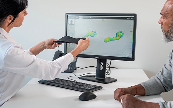
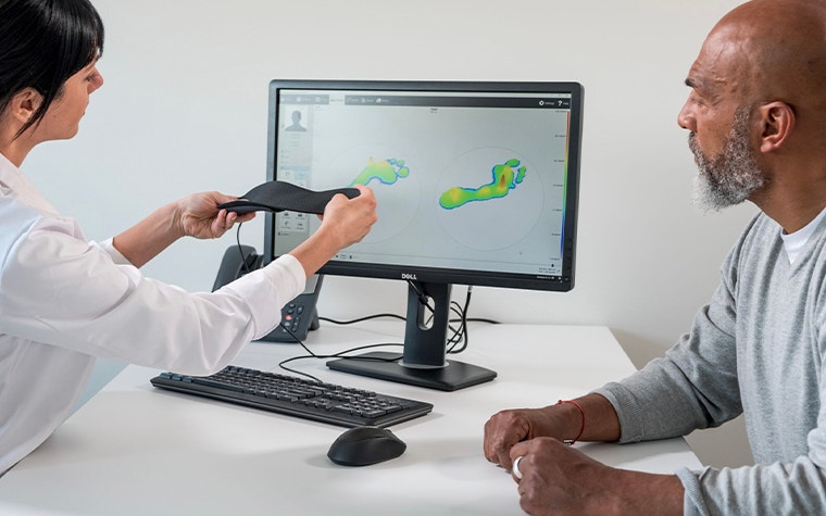
While 2D gait analysis and 3D scanning are both relatively new and advanced technologies supporting this change, they are quite different. 2D scanning offers a comprehensive dynamic pressure measurement of your foot, and 3D scanning provides additional details on the foot's shape, the width of the foot position, and specific anatomical points.
We reached out to Sander Van Nieuwenhoven, Product/Solution Development Specialist at Materialise Motion, to learn more about how the 2D and 3D methods work together to help foot experts achieve better results. Sander, a podiatrist himself, has been intensely involved in developing the Materialise Phits Suite and creating 3D-printed orthotics based on dynamic footscan measurements.
In the past, Materialise Motion (previously RSscan and RS Print) was focused mainly on the 2D dynamic pressure measurement systems. Now, there is more and more integration of the 3D scan. How complementary are the 2D and 3D technologies, and what benefits do we get when combining both methods?
Combining these two methods brings so many benefits. The pressure plate gives us the dynamic information of the patient. We can, for example, see the amount of supination or pronation in the midfoot, the timing of the gait, and the location of the pressure points. This is useful information because it is what we use when deciding the necessary orthotic corrections.
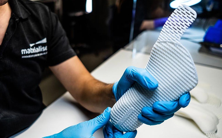
Based on the dynamic information, we recommend the stiffness of the 3D-printed base, which will guide the foot and improve the efficiency of the roll-off. By adding the 3D scan, you get extra information about the height of the arch, the shape of the foot, and so on.
At the moment, these additional digital measurements can help our users better design the orthotics for their patients, for example, by lowering the arch height for patients with a flat foot. Also, the 3D scans help improve communication with patients as we can share the visual information with the patients when explaining their foot morphology.
What are the benefits of having a 3D scan other than having a more detailed and factual visualization?
It brings benefits both to the visualization and the design process not only in terms of additional information but also regarding efficiency. Because it is made digitally, you can add it into your software and combine it with other patient data, like the pressure measurements. Before, clinicians would need additional software to save their 3D scans, but now we combine both 2D and 3D scans in the footscan software.
“Combined with 2D data, 3D scan provides that extra information, which you can use to alter the footscan software's recommendation and modify the dynamic corrections. ”
— Sander Van Nieuwenhoven, Product/Solution Development Specialist, Materialise Motion
Can you be more specific about the parameters a clinician can get from that type of visualization?
The 3D scan gives measurements of the entire foot, like the foot length, foot width, navicular height, arch length, the circumference of the ball of the foot, and more. This data is valuable and related to the entire design of the shoe, not only on the orthotic design. Combined with 2D, 3D scans provide extra information, which you can use to alter the footscan software's recommendation and modify the dynamic recommendation. By adding 3D information into our software, we can already see the benefits because we can adjust the orthotics to suit the patient better.
Can you explain how 2D technology works and what the advantages of using footscan pressure plates are?
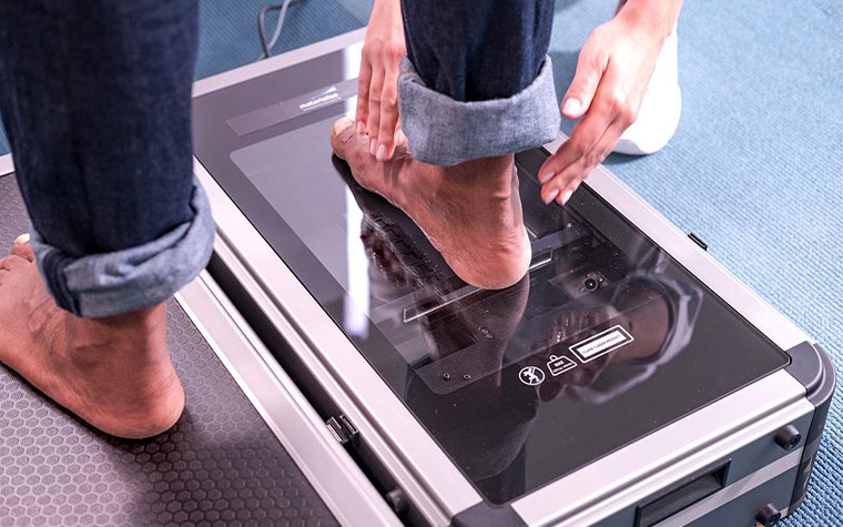
When using our gait analysis system, the patient walks over the footscan pressure plates several times. With their compact sensor density based on three sensors per cm², each analysis is done swiftly and with optimal accuracy. The system captures the patient’s movements and gives us information on the roll-off pattern of the patient. We can look at the complete foot or specific areas. The software divides the foot into 10 anatomical zones and gives you, as a clinician, valuable information on the loading of each of these zones. The system also compares left and right feet. Another great advantage is that you measure movement for a longer time. To extract accurate dynamic measurements, it is crucial that a patient walks several times over the footscan plate to have data to work on.
On the market, there are many 3D scanners to choose from. Can you give us a brief overview of Motion’s scanners and highlight key differences?
Within our Motion portfolio, we offer three types. A small version, iQube mini, has one camera which takes a picture from the bottom of the foot. Then we have a medium version, iQube E500, which has five cameras that capture the sides of the foot to analyze the shape of the arch and the heel. Lastly, we also offer our Tiger 3D scanner, which can measure up to 15 centimeters thanks to its nine cameras.
Is the footscan software compatible with 3D scans taken by devices outside of our portfolio?
Yes. A couple of months ago, we integrated a 3D scan import function that allows you to import any STL or OBJ file from any 3D scanner into the footscan software for expanded analysis.
For example, mobile scanners are very popular at this moment. They are handheld scanners where your patient is sitting, and you go over the foot with the scanner. Now you can upload these 3D scans into our software.
Once a clinician measures and analyzes the patient's foot, what is the next step in finalizing the design and preparing it for manufacturing?
Once you’ve gathered the necessary measurements, the software will calculate the average in the background. On that average, evidence-based algorithms do certain calculations, such as analyzing the balance at specific timings and comparing different loadings. The interesting thing about the footscan software is that it allows users to either follow these recommendations or overrule them, leaving it up to the clinician to decide. Next to the calculated corrections, the user can also manually add other corrections, like metatarsal or forefoot corrections, for example.
And what about the traditional ways of making orthotics, like casting plaster, foam, etc.? How does 2D dynamic scanning compare to those methods?
The traditional methods for making orthotics are often messy and require more time to take the measurements and make the product. The result is also very dependent on the person who makes the soles and at what time, as it is manual work. The use of a pressure plate, together with a digital workflow, solves these problems as your patient just has to walk or run over the platform to get their measurements. Also, because you will have multiple foot strikes per patient, you will have a much more accurate impression of the patient’s feet, than when you would only take a static copy of the foot.
While the traditional techniques take up to 30 to 40 minutes to design a pair of insoles, 2D gait analysis takes only 15 to 20 minutes, cutting the required test and design time in half. That means that a clinician can see more patients daily because it takes less time to examine each patient. The Materialise Phits Suite workflow also makes it easy and fast to duplicate orthotics.
How can foot experts use footscan to better explain to their patient the treatment and the benefits of the phits orthotics?
The footscan software is very intuitive. Data retrieved from pressure plates allows you to explain to the patient the issues with their gait. Quite often, you can link complaints to specific patterns. For example, if a patient has pain in his forefoot, you will be able to show them a red high-pressure area on their forefoot. Also, it’s great to share the information afterward with their general practitioner. They can use it in the future diagnosis, and the patients can use the insights to buy specific shoes, especially for sports.
Can you explain the research that goes into designing the footscan software?
At Motion, research is fundamental. We participate in extensive research projects with other organizations and universities and gather valuable data on shoes, orthotics, foot morphology, movement, and their connections. Guided by scientific studies, we continuously improve our algorithms and make them more accurate. Our data-driven approach guides us in optimizing our offer and developing future-proof solutions.
Two research studies are particularly important. Thanks to them, we know it is possible to measure the risk of your patient getting an injury.
In the first study, subjects were divided into three risk groups based on their roll-off pattern. The results showed that the subjects of the medium and the high-risk groups had significantly more lower leg injuries than the subjects from the low-risk group.
In the second study, custom orthotics were added as a preventive solution. We divided the medium- and high-risk groups in half: one group got custom insoles based on the current phits algorithm, and the other group didn't. The subject group — with insoles — had significantly less injuries than the other subject group — without insoles. The conclusion is that the footscan software can detect a patient's risk of getting an injury, and we can reduce the injury rate by providing custom insoles to medium and high-risk patients.
What is coming next for Materialise Motion? What are the objectives you want to achieve in the future?
We will proceed with developing and combining both techniques to use the best of both worlds. The goal is to use the dynamic information while still considering the foot's shape and through that powerful synergy, create the best possible orthotics that will help people improve their movement and quality of life.
Share on:
You might also like
Never miss a story like this. Get curated content delivered straight to your inbox.