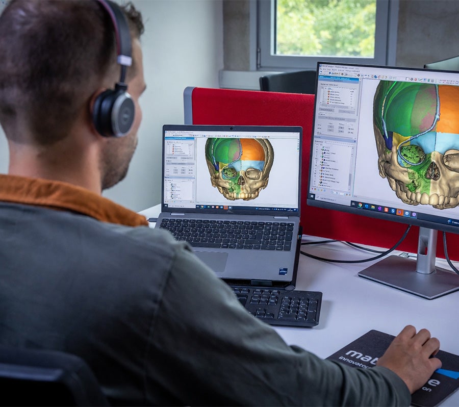
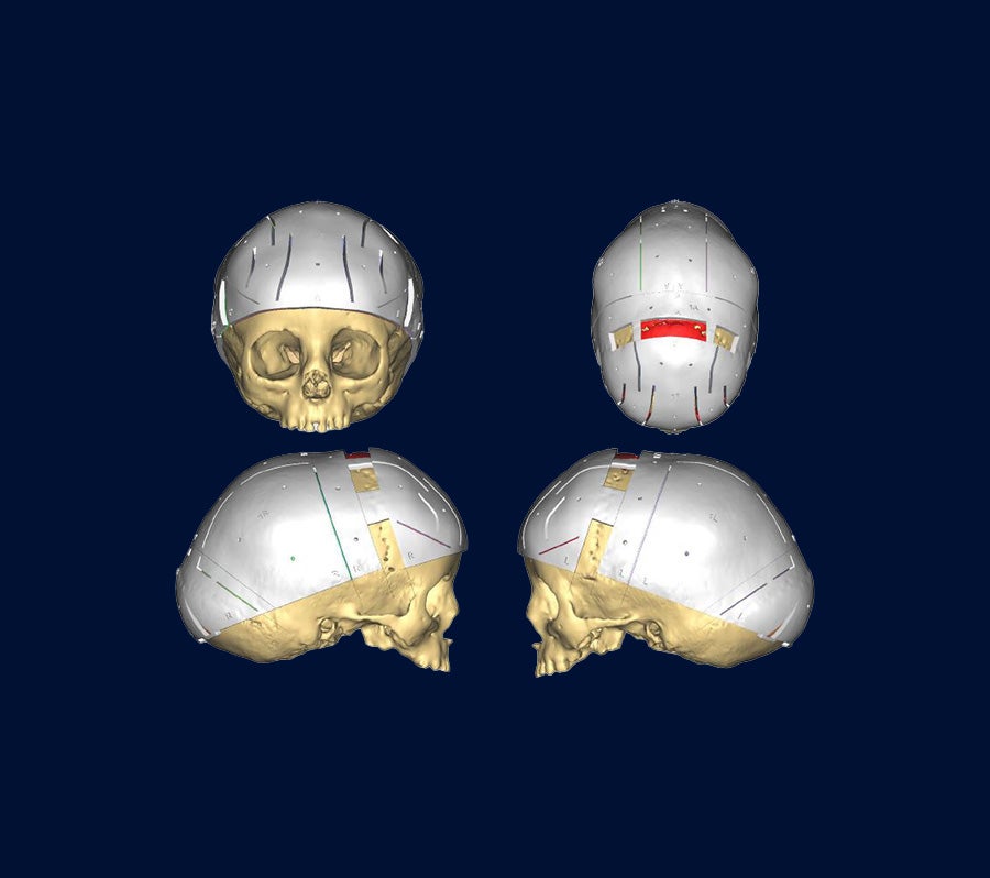
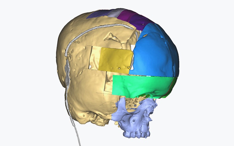
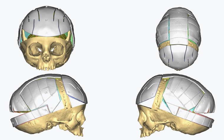
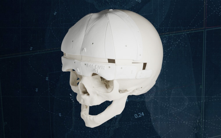
Cranial Vault Reconstruction for Craniosynostosis

Predictability, planned in 3D

When the most delicate patients are under anesthesia, every second counts. Make surgical decisions in advance with 3D planning to spend OR time operating, not deliberating. Our clinical engineers closely collaborate with you to translate your preferred cranial vault reconstruction technique into precise 3D tools for objective, data-driven surgical preparation.

A 3D-printed anatomical model of an infant human skull with cranial cutting guides, displayed against a dark background.

More operating, less deliberating

Define the ideal outcome before the first incision

Rely on surgical tools from your unique plan

Operate with confidence and efficiency

Close the loop on quality and learning

For cranial vault reconstruction, 3D planning combined with patient-specific guides is now our routine approach in Denmark. It allows us to define the surgical plan in detail beforehand and translate it accurately into the operating room.

Headshot of Dr. Thomas Kofod, Chair of the Department of Oral and Maxillofacial Surgery at the Rigshospital in Denmark
Dr. Thomas KofodConsultant, Department of Oral and Maxillofacial Surgery Rigshospitalet, Copenhagen, Denmark

With a full 3D plan and patient-specific guides, I walk into the OR knowing exactly where we’ll cut, how we will reposition, and the symmetry we’ll achieve. It takes the uncertainty out of high-stakes cases and lets me focus on execution.

Dr. Antonio Huete, MDNeurosurgeon, Department of CMF — Pediatric Craniofacial Unit Hospital Universitario Torrecárdenas, Spain

With virtual 3D planning and patient-specific guides, we can perform the surgery before entering the operating room. It transforms complex cranial vault reconstruction into a predictable, controlled procedure.

Dr. Juana VidalPediatric CMF Surgeon, Reina Sofía University Hospital

Your tools for predictable outcomes

Plan in 3D with advanced tools

  • Segment critical anatomy, such as the sagittal and transverse sinuses, to plan safely and avoid intraoperative risk
  • Use patient-specific mirroring to define the optimal cranial shape and balance symmetry with volume expansion
  • Account for constraints like previous surgeries or implants directly in your 3D plan
Digital rendering of an infant human skull with multicolored segmented cranial implant sections in blue, green, yellow, and purple.

Perform the operation with personalized surgical guides and shaping templates

  • Use patient-specific cutting guides to accurately transfer planned osteotomies to the surgical field
  • Match fixation and the final shape to your plan with repositioning guides
  • Minimize intraoperative uncertainty so you can concentrate on execution, not recalculation
Four angles of an infant skull model with personalized positioning guides attached.

Gain clarity with 3D-printed anatomical models and XR visualization

  • 3D print physical anatomical models for tactile OR reference, teaching, and family communication
  • Visualize your plan in extended reality (XR) to build immersive spatial understanding of the anatomy and planned procedure
  • Rehearse the operation in advance to align the surgical team on the approach
A surgeon wearing a head-mounted augmented reality Microsoft Hololens device gestures while working in an operating room with medical equipment in the background.

Delivering precision at Reina Sofía University Hospital