
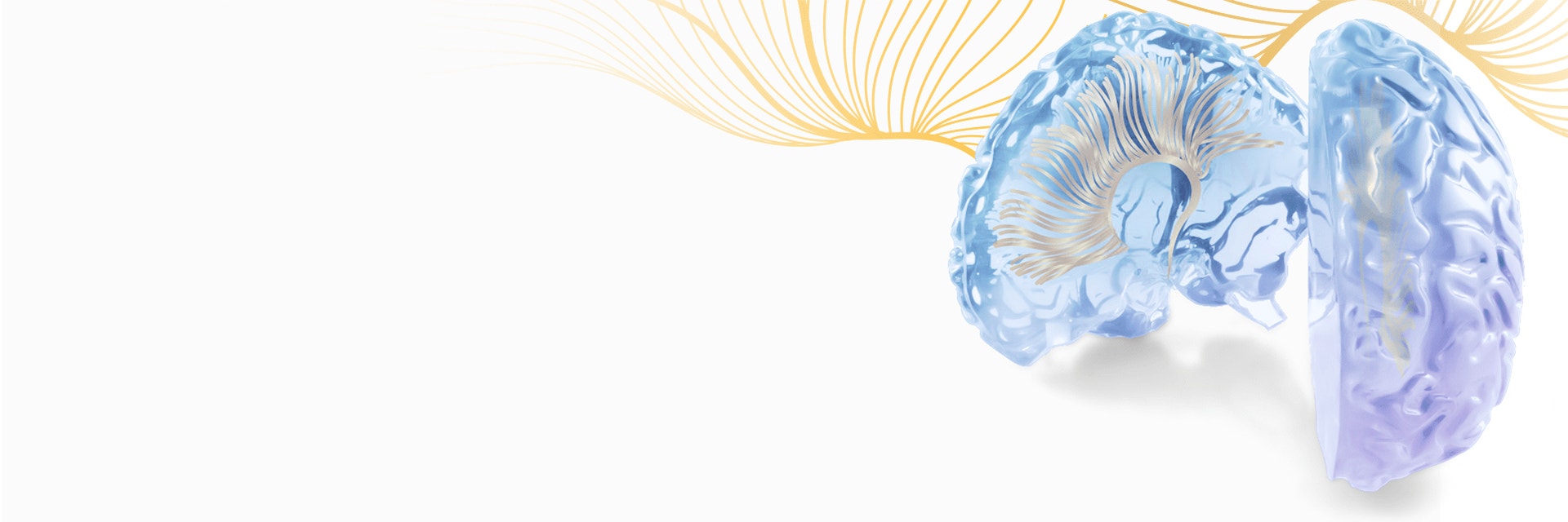
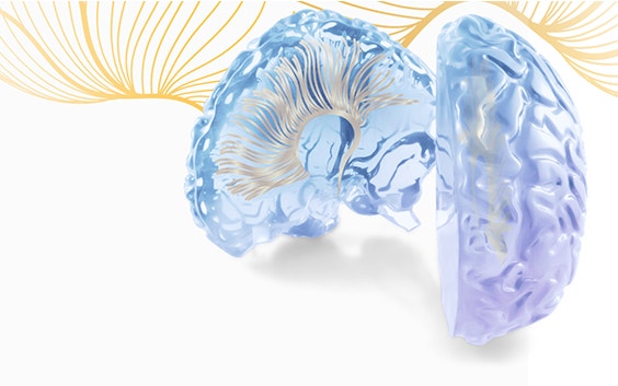
EXPERT INSIGHT
Novel Insights About Dissecting Abdominal Aortic Aneurysms in Mice
2 min read
Abdominal aortic aneurysms (AAA) occur in 5 to 9% of the population over the age of 65 years and transmural aneurysm rupture is the 10th most common cause of death in the industrialized world. Dr. Bram Trachet, post-doctoral researcher at École Polytechnique Fédérale de Lausanne (EPFL) in Switzerland, explores novel high-resolution imaging techniques as well as image-guided histology to visualize experimental aneurysms in laboratory animals.
In 2015, he won a Mimics Innovation Award for his research on the morphology of abdominal aortic aneurysms in mice infused with angiotensin II. The essence of his paper is presented in this blog post.

Of mice and men
Although mice are the most commonly used animals in aneurysm research, there are some important differences between aneurysms that form in mice treated with angiotensin II, on the one hand, and aneurysms that form spontaneously in humans, on the other hand. In mice, the abdominal aortic aneurysm forms suprarenally (i.e. above the kidneys), while in humans most aneurysms form infrarenally (i.e. below the kidneys). Luminal dilation also develops suddenly in mice instead of growing progressively. Aneurysms in mice often develop an intramural thrombus rather than an intraluminal thrombus as we see in human aneurysms.
While the mouse model has been used to study aneurysms in over 200 papers, these morphological differences have never been explained. Dr. Trachet posited that better imaging techniques (such as visualizing the aortic wall in high-resolution 3D) might allow him to gain a deeper understanding of the processes that led to these phenomena.
Getting a 3D optimized model of abdominal aortic aneurysms
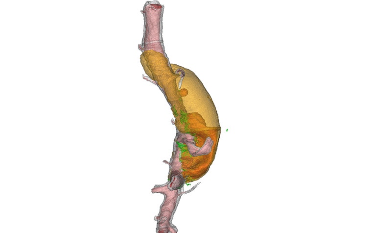
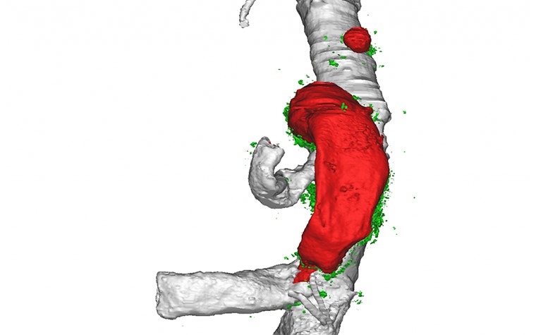
The 3D reconstruction of the aortic wall proved to be challenging, as the conventional methods of ultrasound and in vivo micro-CT provided images which were not sufficiently detailed for a 3D model of the aortic wall. In vivo micro-MRI could allow for a better soft tissue contrast, but the through-plane distance is too coarse for a detailed 3D analysis. In the end, differential phase contrast X-ray tomographic microscopy (PCXTM) was used, as it uses synchrotron radiation to overcome all the limitations of the above methods. All reconstructed datasets (in vivo as well as PCXTM) were then semi-automatically segmented into 3D models using Materialise Mimics Research software.
A new insight
The virtual 3D models greatly enhanced Dr. Trachet's capacity to study the morphology of the abdominal aortic aneurysms present in angiotensin II-treated mice. Due to these new imaging techniques, the exact culprit for the lesions could be determined for the first time ever. It turned out that small suprarenal side branches, not visible using traditional imaging techniques, were responsible for the intramural hematoma, and therefore were also responsible for the suprarenal location of the aneurysms. Moreover, as Dr. Trachet found out from his new and improved view of the aortic wall, wherever a luminal expansion appeared on histology, it was invariably accompanied by a tear in the tunica media. This explained the sudden dilatation: aneurysms in these mice are more reminiscent of aortic dissections. In conclusion, his research was able to expand the existing paradigm, providing answers to questions that had lingered for many years.
L-102651
Share on:

Meet the author
Dr. Bram Trachet
You might also like
Never miss a story like this. Get curated content delivered straight to your inbox.