CUSTOMER STORY
3D-Printed Kidney Model Allows Surgeons to Successfully Transplant Adult Kidney into Two-Year-Old
3D-printed anatomical models for diagnostic use created with MIS/Mimics are not commercially available in the US, Australia and Canada.
For the first time ever, surgeons at Guy’s and St. Thomas’ NHS Foundation Trust have been able to transplant an adult kidney into a two-year-old child, using 3D printing to achieve this complex surgery.
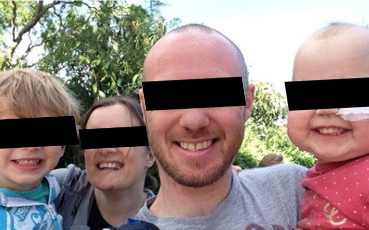

Two-year-old Lucy Boucher was the victim of supraventricular tachycardia as a baby, which resulted in heart failure. Her body was starved of oxygen, which in turn caused a kidney failure. The outlook was bleak: although Lucy had undergone corrective heart surgery, she looked set to spend the rest of her life undergoing dialysis treatment.
The situation changed when Lucy was referred to the Guy’s and St. Thomas’ NHS Foundation Trust in London. The surgeons at the hospital suggested a new possibility: that Lucy’s father could replace his daughter’s kidneys with one of his own. But this posed the problem of fitting an adult-sized kidney into the abdomen of a small child, and before Lucy’s doctors began the surgery they needed to have an exact idea of how to position the organ.
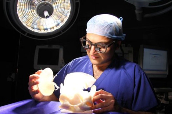

Due to the complexity of the case, the surgical team would need to rely on a multidisciplinary approach. And the key to uniting experts in various disciplines? 3D Printing. Surgeons usually rely on radiologists to give them a better understanding of their patients’ anatomies with x-rays and scans – but in more complex cases such as Lucy’s, it was important to visualize the complex operation in 3D to give the most accurate impression of her anatomy.
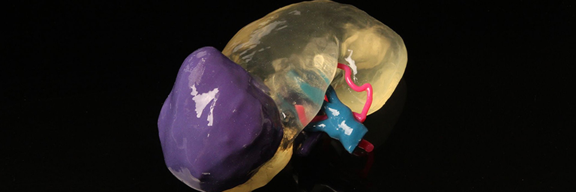
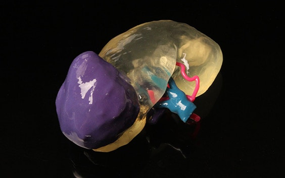
Lucy’s abdomen was scanned and segmented into Materialise Mimics®, and recreated in 3D along with crucial parts of her anatomy such as the aorta, the coeliac axis, the superior mesenteric artery, the common iliac arteries, the inferior vena cava, the common iliac veins, pelvic bones, the lateral abdominal wall, and the inferior margin. Then her father’s kidney was also scanned and turned into a 3D model. Finally, both models were sent off to the 3D printer at the Guy’s and St. Thomas’ 3D Printing Facility — the hospital works with PolyJet technology, which allows them to create 3D medical models in different materials in order to replicate the different mechanical properties of different sorts of tissues.
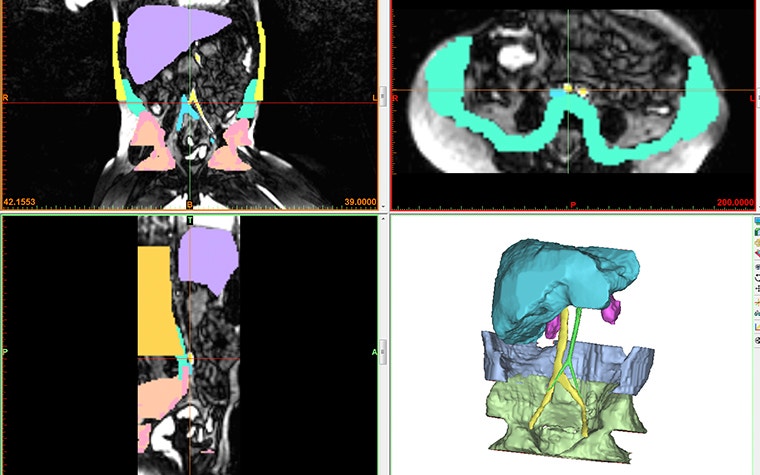

The 3D-printed kidney models were crucial in giving the surgeons a clear idea of how to do the surgery before even making the first cut. The anatomical model allowed them to plan the incision, how to connect the kidney to the blood vessels and the respective anastomosis sites, as well as how to position the organ within the abdomen. As Dr. Pankaj Chandak states, “The most important benefit is to patient safety. The 3D-printed models allow informative, hands-on planning, ahead of the surgery with replicas that are the next best thing to the actual organs themselves. This means surgeons are better placed than before to prepare for the operation and to assess what surgical approach will offer the greatest chance of a safe and successful transplant.”
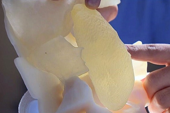

And not only the surgeons benefit from this approach; Lucy’s parents were able to clearly understand what was about to happen to their daughter. Lucy’s father Chris explained that “Seeing the model of her abdomen and the way the kidney was going to be transplanted inside her gave me a clear understanding of exactly what was going to happen. It helped ease my concerns and it was hugely reassuring to know that the surgeons could carry out such detailed planning ahead of the operation.”
As 3D printing becomes increasingly accessible to hospitals, 3D-printed anatomical models are becoming a common feature in hospitals. Today clinicians can create their anatomical models in a few clicks using Mimics® inPrint.
以下で共有する:
