EXPERT INSIGHT
Patient-Specific 3D Modeling: An Innovative Approach for Scoliosis Diagnosis
It's a bright new day in Serbia. Researchers at the University of Kragujevac have proposed a new, non-invasive 3D methodology for assessing scoliosis, which allows for spine measurements to be performed while limiting radiation exposure and human intervention.
Scoliosis in young patients
6 – 8% of the world's population is affected by scoliosis. It is a very complex spinal disorder, and children and adolescents make up the most vulnerable group. Reducing radiation exposure, or eliminating it altogether as an assessment method, is imperative
A need for new diagnostic techniques
In scoliosis, side-to-side curvature of the spine is common, typically involving rotation of the vertebrae. Usually, the disorder is examined and diagnosed with some form of radiation, such as CT or X-ray. This can result in harmful side effects for the patients.
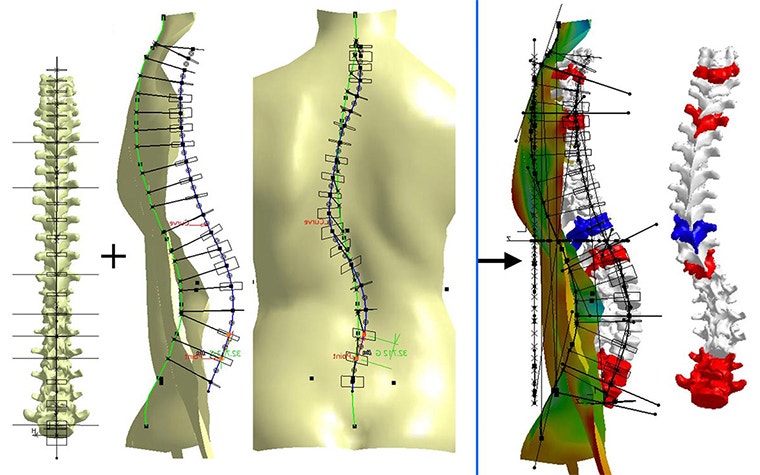
Current scoliosis diagnostic methods have other drawbacks. The 2D nature of a planar X-ray and the fact that these CT-based methods do not offer a standing position of the patient's spine can limit the accuracy and detailing of the assessment.
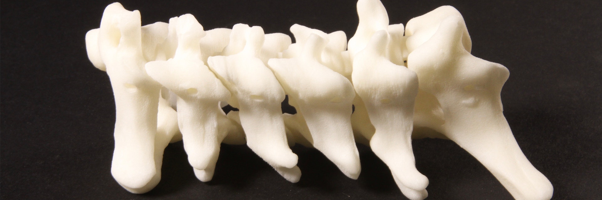
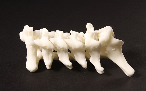
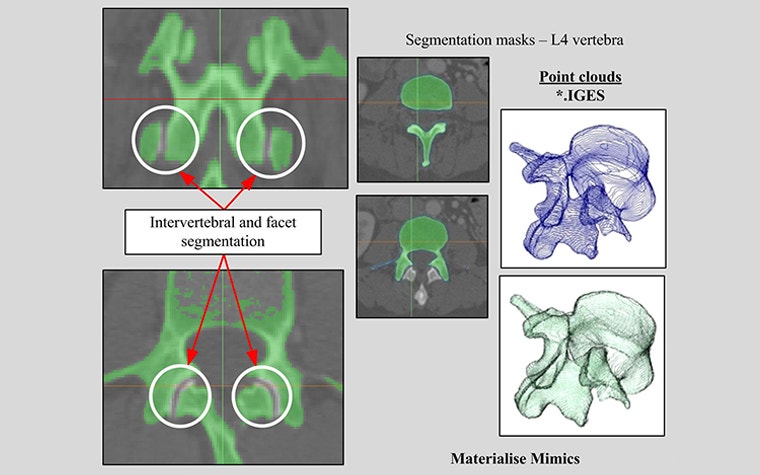
Materialise Mimics for 3D spinal reconstruction
The research in the University of Kragujevac's published paper is based on a target group of 372 patients with various types of spinal deformity. In their research, the university used Mimics' segmentation algorithms for the 3D spinal reconstruction of CT images, and combined it with dorsal optical scans to generate a model of the spinal deformity. This provides a more comprehensive analysis of the disorder.
It's been proven that the system used by the university demonstrates a reduction in exposure to radiation by replacing X-ray-based scans with optical scans, while still allowing for 3D measurements. As the 3D nature of the deformity is not paid enough attention in daily clinical practice, researchers expect that this new protocol will show its high value when these 3D measurements are performed on scoliosis patients.
Working towards eliminating radiation
Non-invasive, accurate, and faster scoliosis diagnosis methods may be making their way into the medical world faster than we think. Their advantages show obvious promise and give us a positive indication that patient-specific 3D modeling could be a step closer to eliminating X-ray radiation and improving patient diagnoses.
Share on:
You might also like
Never miss a story like this. Get curated content delivered straight to your inbox.