PATIENT STORY
Baby Born with Brain outside His Skull Saved by Surgeons at Boston Children’s Hospital
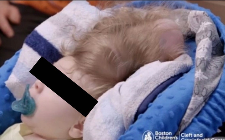
Thanks to medical 3D printing, surgeons have treated a young baby with an encephalocele: a rare disorder where part of the fetus’s brain starts growing outside its head in the womb.
Dr. John Meara from the Cleft and Craniofacial Center at Boston Children’s was able to carry out this extremely difficult procedure thanks to thorough preparation in collaboration with the hospital’s Simulator Program (SIMPeds), which enables doctors to rehearse their procedures via a variety of methods, including 3D printing. 3D printing plays a large role in providing the surgeons with tangible anatomical models they can use to plan their surgery. Materialise has been proud to support the SIMPeds program by sharing its state-of-the-art expertise in 3D printing.
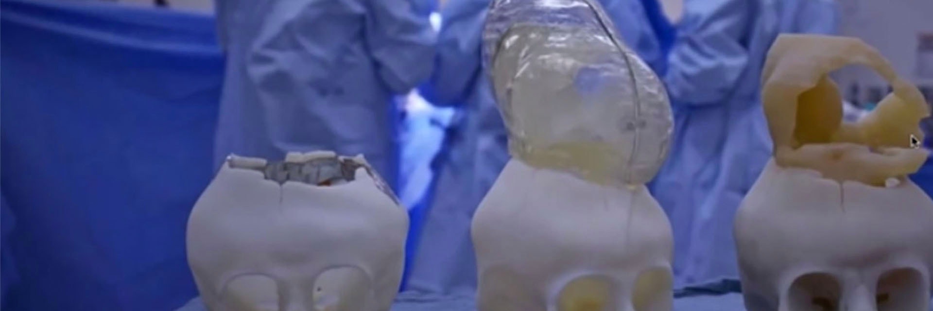
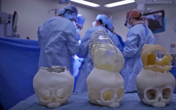
Using a CT scan of the baby’s skull, the SIMPeds technicians were able to create a 3D-printed model for Dr. Meara and his colleague, Dr. Mark Proctor, to allow for a more tangible pre-surgical planning. Another added benefit of printing out the skull was that the surgeons could clearly explain the operation to the baby’s parents and thereby provide a measure of reassurance.
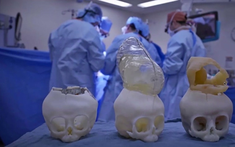
The biggest obstacle in the surgery was the size of the baby’s skull — it was too small to fit the entire brain inside it, so Dr. Meara and Dr. Proctor came up with the idea of making a series of incisions in the bone that would allow the skull to expand. The procedure took around five hours in total — which they deemed much shorter than it would have been without the precise planning beforehand — and the surgeons successfully managed to place the brain back inside the skull.
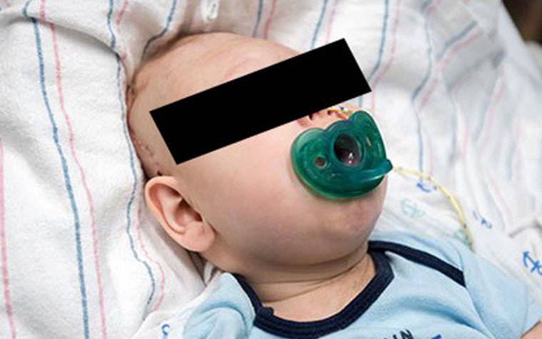
Although the baby recovered well from the operation, it is too early to tell how he will develop. His brain has such a unique structure that doctors have no way of telling how normal his functions will be. But the operation went exactly according to plan, and he has been given the best possible chance at a normal and healthy life!
3D-printed anatomical models for diagnostic use created with Mimics Innovation Suite are not commercially available in the US, Australia, and Canada.
Share on:
You might also like
Never miss a story like this. Get curated content delivered straight to your inbox.